E Radin, A V Marcuzzo, J de Groodt, F Degrassi, L Calderan, V Ramella, G Tirelli, M Ukmar, M A Cova
{"title":"基于核磁共振成像的口腔鳞状细胞癌肌层评估--7点评分法","authors":"E Radin, A V Marcuzzo, J de Groodt, F Degrassi, L Calderan, V Ramella, G Tirelli, M Ukmar, M A Cova","doi":"10.1007/s00330-024-11016-8","DOIUrl":null,"url":null,"abstract":"<p><strong>Objectives: </strong>To investigate preoperative MRI evaluation of the features of the mylohyoid muscle (MM) predictive of its infiltration in oral squamous cell carcinoma (OSCC) treatment planning, defining the most appropriate sequences to study its deep extension into the floor of the mouth (FOM).</p><p><strong>Materials and methods: </strong>We applied a 7-point score to retrospectively evaluate preoperative imaging of patients who underwent surgery for OSCC over 11 years. The results were compared with histopathological findings using Spearman's rank coefficient. Receiver operating characteristic curves were employed to assess the score's ability to predict MM infiltration, determining optimal thresholds for sensitivity, specificity, and predictive values. The Mann-Whitney U-test confirmed that infiltration judgments did not overlap around this threshold. Cohen's K statistical coefficient was used to evaluate the interobserver agreement.</p><p><strong>Results: </strong>Fifty-two patients (mean age 66.4 ± 11.9 years, 36 men) were evaluated. Histopathological examination found MM infiltration in 21% of cases (n = 11), with 90% classified in the highest Score categories. A score > 4 proved to be the best cut-off for predicting the risk of MM infiltration, with a sensitivity of 91% (CI: 0.57-0.99), specificity 61% (CI: 0.45-0.76), PPV 38% (CI: 0.21-0.59), and NPV 96% (CI: 0.78-0.99). At the subsequent single-sequence assessment, the TSE-T2wi had the highest diagnostic accuracy, with sensitivity 90% (CI: 0.57-0.99), specificity 70% (CI: 0.53-0.82), PPV 45% (CI: 0.25-0.67), and NPV 96% (CI: 0.80-0.99).</p><p><strong>Conclusion: </strong>The 7-point score is a promising predictor of safe surgical margins for MM in OSCC treatment, with the particular benefit of T2-weighted sequences.</p><p><strong>Clinical relevance statement: </strong>Our scoring system for tumor infiltration of MM, which is easy to use even for less experienced radiologists, allows for uniformity in radiological language, thereby ensuring crucial preoperative information for the surgeon.</p><p><strong>Key points: </strong>The relationship of the MM to an oral lesion may impact surgical planning. As the score increases, there is a greater incidence of infiltration in the MM. Our score system improves radiologists' reporting for MM involvement by tumor.</p>","PeriodicalId":12076,"journal":{"name":"European Radiology","volume":" ","pages":"2065-2073"},"PeriodicalIF":4.7000,"publicationDate":"2025-04-01","publicationTypes":"Journal Article","fieldsOfStudy":null,"isOpenAccess":false,"openAccessPdf":"https://www.ncbi.nlm.nih.gov/pmc/articles/PMC11913961/pdf/","citationCount":"0","resultStr":"{\"title\":\"MRI-based assessment of the mylohyoid muscle in oral squamous cell carcinoma, a 7-point scoring method.\",\"authors\":\"E Radin, A V Marcuzzo, J de Groodt, F Degrassi, L Calderan, V Ramella, G Tirelli, M Ukmar, M A Cova\",\"doi\":\"10.1007/s00330-024-11016-8\",\"DOIUrl\":null,\"url\":null,\"abstract\":\"<p><strong>Objectives: </strong>To investigate preoperative MRI evaluation of the features of the mylohyoid muscle (MM) predictive of its infiltration in oral squamous cell carcinoma (OSCC) treatment planning, defining the most appropriate sequences to study its deep extension into the floor of the mouth (FOM).</p><p><strong>Materials and methods: </strong>We applied a 7-point score to retrospectively evaluate preoperative imaging of patients who underwent surgery for OSCC over 11 years. The results were compared with histopathological findings using Spearman's rank coefficient. Receiver operating characteristic curves were employed to assess the score's ability to predict MM infiltration, determining optimal thresholds for sensitivity, specificity, and predictive values. The Mann-Whitney U-test confirmed that infiltration judgments did not overlap around this threshold. Cohen's K statistical coefficient was used to evaluate the interobserver agreement.</p><p><strong>Results: </strong>Fifty-two patients (mean age 66.4 ± 11.9 years, 36 men) were evaluated. Histopathological examination found MM infiltration in 21% of cases (n = 11), with 90% classified in the highest Score categories. A score > 4 proved to be the best cut-off for predicting the risk of MM infiltration, with a sensitivity of 91% (CI: 0.57-0.99), specificity 61% (CI: 0.45-0.76), PPV 38% (CI: 0.21-0.59), and NPV 96% (CI: 0.78-0.99). At the subsequent single-sequence assessment, the TSE-T2wi had the highest diagnostic accuracy, with sensitivity 90% (CI: 0.57-0.99), specificity 70% (CI: 0.53-0.82), PPV 45% (CI: 0.25-0.67), and NPV 96% (CI: 0.80-0.99).</p><p><strong>Conclusion: </strong>The 7-point score is a promising predictor of safe surgical margins for MM in OSCC treatment, with the particular benefit of T2-weighted sequences.</p><p><strong>Clinical relevance statement: </strong>Our scoring system for tumor infiltration of MM, which is easy to use even for less experienced radiologists, allows for uniformity in radiological language, thereby ensuring crucial preoperative information for the surgeon.</p><p><strong>Key points: </strong>The relationship of the MM to an oral lesion may impact surgical planning. As the score increases, there is a greater incidence of infiltration in the MM. Our score system improves radiologists' reporting for MM involvement by tumor.</p>\",\"PeriodicalId\":12076,\"journal\":{\"name\":\"European Radiology\",\"volume\":\" \",\"pages\":\"2065-2073\"},\"PeriodicalIF\":4.7000,\"publicationDate\":\"2025-04-01\",\"publicationTypes\":\"Journal Article\",\"fieldsOfStudy\":null,\"isOpenAccess\":false,\"openAccessPdf\":\"https://www.ncbi.nlm.nih.gov/pmc/articles/PMC11913961/pdf/\",\"citationCount\":\"0\",\"resultStr\":null,\"platform\":\"Semanticscholar\",\"paperid\":null,\"PeriodicalName\":\"European Radiology\",\"FirstCategoryId\":\"3\",\"ListUrlMain\":\"https://doi.org/10.1007/s00330-024-11016-8\",\"RegionNum\":2,\"RegionCategory\":\"医学\",\"ArticlePicture\":[],\"TitleCN\":null,\"AbstractTextCN\":null,\"PMCID\":null,\"EPubDate\":\"2024/8/29 0:00:00\",\"PubModel\":\"Epub\",\"JCR\":\"Q1\",\"JCRName\":\"RADIOLOGY, NUCLEAR MEDICINE & MEDICAL IMAGING\",\"Score\":null,\"Total\":0}","platform":"Semanticscholar","paperid":null,"PeriodicalName":"European Radiology","FirstCategoryId":"3","ListUrlMain":"https://doi.org/10.1007/s00330-024-11016-8","RegionNum":2,"RegionCategory":"医学","ArticlePicture":[],"TitleCN":null,"AbstractTextCN":null,"PMCID":null,"EPubDate":"2024/8/29 0:00:00","PubModel":"Epub","JCR":"Q1","JCRName":"RADIOLOGY, NUCLEAR MEDICINE & MEDICAL IMAGING","Score":null,"Total":0}
引用次数: 0
摘要
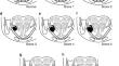
目的研究在口腔鳞状细胞癌(OSCC)治疗计划中,术前核磁共振成像(MRI)评估可预测其浸润的舌骨肌(MM)特征,确定研究其向口底(FOM)深部延伸的最合适序列:我们采用 7 点评分法对 11 年来接受手术治疗的 OSCC 患者的术前成像进行了回顾性评估。使用斯皮尔曼秩系数将结果与组织病理学结果进行比较。采用接收者操作特征曲线评估该评分预测 MM 浸润的能力,确定敏感性、特异性和预测值的最佳阈值。曼-惠特尼 U 检验证实,浸润判断在该阈值附近没有重叠。科恩 K 统计系数用于评估观察者之间的一致性:共评估了 52 名患者(平均年龄为 66.4 ± 11.9 岁,36 名男性)。组织病理学检查发现 21% 的病例(n = 11)存在 MM 浸润,90% 的病例被归入最高评分类别。事实证明,得分大于 4 分是预测 MM 浸润风险的最佳临界值,灵敏度为 91%(CI:0.57-0.99),特异性为 61%(CI:0.45-0.76),PPV 为 38%(CI:0.21-0.59),NPV 为 96%(CI:0.78-0.99)。在随后的单序列评估中,TSE-T2wi 的诊断准确性最高,灵敏度为 90%(CI:0.57-0.99),特异性为 70%(CI:0.53-0.82),PPV 为 45%(CI:0.25-0.67),NPV 为 96%(CI:0.80-0.99):7点评分是预测OSCC治疗中MM安全手术切缘的有效指标,T2加权序列尤其具有优势:我们的MM肿瘤浸润评分系统即使对经验不足的放射科医生来说也很容易使用,它可以实现放射学语言的统一,从而确保为外科医生提供关键的术前信息:要点:MM 与口腔病变的关系可能会影响手术计划。要点:MM 与口腔病变的关系可能会影响手术计划,随着评分的增加,MM 浸润的发生率也会增加。我们的评分系统提高了放射医师对 MM 受肿瘤累及情况的报告能力。
MRI-based assessment of the mylohyoid muscle in oral squamous cell carcinoma, a 7-point scoring method.
Objectives: To investigate preoperative MRI evaluation of the features of the mylohyoid muscle (MM) predictive of its infiltration in oral squamous cell carcinoma (OSCC) treatment planning, defining the most appropriate sequences to study its deep extension into the floor of the mouth (FOM).
Materials and methods: We applied a 7-point score to retrospectively evaluate preoperative imaging of patients who underwent surgery for OSCC over 11 years. The results were compared with histopathological findings using Spearman's rank coefficient. Receiver operating characteristic curves were employed to assess the score's ability to predict MM infiltration, determining optimal thresholds for sensitivity, specificity, and predictive values. The Mann-Whitney U-test confirmed that infiltration judgments did not overlap around this threshold. Cohen's K statistical coefficient was used to evaluate the interobserver agreement.
Results: Fifty-two patients (mean age 66.4 ± 11.9 years, 36 men) were evaluated. Histopathological examination found MM infiltration in 21% of cases (n = 11), with 90% classified in the highest Score categories. A score > 4 proved to be the best cut-off for predicting the risk of MM infiltration, with a sensitivity of 91% (CI: 0.57-0.99), specificity 61% (CI: 0.45-0.76), PPV 38% (CI: 0.21-0.59), and NPV 96% (CI: 0.78-0.99). At the subsequent single-sequence assessment, the TSE-T2wi had the highest diagnostic accuracy, with sensitivity 90% (CI: 0.57-0.99), specificity 70% (CI: 0.53-0.82), PPV 45% (CI: 0.25-0.67), and NPV 96% (CI: 0.80-0.99).
Conclusion: The 7-point score is a promising predictor of safe surgical margins for MM in OSCC treatment, with the particular benefit of T2-weighted sequences.
Clinical relevance statement: Our scoring system for tumor infiltration of MM, which is easy to use even for less experienced radiologists, allows for uniformity in radiological language, thereby ensuring crucial preoperative information for the surgeon.
Key points: The relationship of the MM to an oral lesion may impact surgical planning. As the score increases, there is a greater incidence of infiltration in the MM. Our score system improves radiologists' reporting for MM involvement by tumor.
期刊介绍:
European Radiology (ER) continuously updates scientific knowledge in radiology by publication of strong original articles and state-of-the-art reviews written by leading radiologists. A well balanced combination of review articles, original papers, short communications from European radiological congresses and information on society matters makes ER an indispensable source for current information in this field.
This is the Journal of the European Society of Radiology, and the official journal of a number of societies.
From 2004-2008 supplements to European Radiology were published under its companion, European Radiology Supplements, ISSN 1613-3749.

 求助内容:
求助内容: 应助结果提醒方式:


