Tahani Ahmad, Alessandro Guida, Samuel Stewart, Noah Barrett, Xiang Jiang, Michael Vincer, Jehier Afifi
{"title":"深度学习能否对脑超声图像进行分类,以检测极早产儿的脑损伤?","authors":"Tahani Ahmad, Alessandro Guida, Samuel Stewart, Noah Barrett, Xiang Jiang, Michael Vincer, Jehier Afifi","doi":"10.1007/s00330-024-11028-4","DOIUrl":null,"url":null,"abstract":"<p><strong>Objectives: </strong>Cerebral ultrasound (CUS) is the main imaging screening tool in preterm infants. The aim of this work is to develop deep learning (DL) models that classify normal vs abnormal CUS to serve as a computer-aided detection tool providing timely interpretation of the scans.</p><p><strong>Methods: </strong>A population-based cohort of very preterm infants (22<sup>0</sup>-30<sup>6</sup> weeks) born between 2004 and 2016 in Nova Scotia, Canada. A set of nine sequential CUS images per infant was retrieved at three specific coronal landmarks at three pre-identified times (first, sixth weeks, and term age). A radiologist manually labeled each image as normal or abnormal. The dataset was split into training/development/test subsets (80:10:10). Different convolutional neural networks were tested, with filtering of the most uncertain prediction. The model's performance was assessed using precision/recall and the receiver operating area under the curve.</p><p><strong>Results: </strong>Sequential CUS retrieved for 538/665 babies (81% of the cohort). Four thousand one hundred eighty images were used to develop and test the model. The model performance was only discrete at the beginning but, through different machine learning strategies was boosted to good levels averaging 0.86 ROC AUC (95% CI: 0.82, 0.90) and 0.87 PR AUC (95% CI: 0.84, 0.90) (model uncertainty estimation filters using normalized entropy threshold = 0.5).</p><p><strong>Conclusion: </strong>This study offers proof of the feasibility of applying DL to CUS. This basic diagnostic model showed good discriminative ability to classify normal versus abnormal CUS. This serves as a CAD and a framework for constructing a prognostic model.</p><p><strong>Clinical relevance statement: </strong>This DL model can serve as a computer-aided detection tool to classify CUS of very preterm babies as either normal or abnormal. This model will also be used as a framework to develop a prognostic model.</p><p><strong>Key points: </strong>Binary computer-aided detection models of CUS are applicable for classifying ultrasound images in very preterm babies. This model acts as a step towards developing a model for predicting neurodevelopmental outcomes in very preterm babies. This model serves as a tool for interpretation of CUS in this patient population with a heightened risk of brain injury.</p>","PeriodicalId":12076,"journal":{"name":"European Radiology","volume":" ","pages":"1948-1958"},"PeriodicalIF":4.7000,"publicationDate":"2025-04-01","publicationTypes":"Journal Article","fieldsOfStudy":null,"isOpenAccess":false,"openAccessPdf":"","citationCount":"0","resultStr":"{\"title\":\"Can deep learning classify cerebral ultrasound images for the detection of brain injury in very preterm infants?\",\"authors\":\"Tahani Ahmad, Alessandro Guida, Samuel Stewart, Noah Barrett, Xiang Jiang, Michael Vincer, Jehier Afifi\",\"doi\":\"10.1007/s00330-024-11028-4\",\"DOIUrl\":null,\"url\":null,\"abstract\":\"<p><strong>Objectives: </strong>Cerebral ultrasound (CUS) is the main imaging screening tool in preterm infants. The aim of this work is to develop deep learning (DL) models that classify normal vs abnormal CUS to serve as a computer-aided detection tool providing timely interpretation of the scans.</p><p><strong>Methods: </strong>A population-based cohort of very preterm infants (22<sup>0</sup>-30<sup>6</sup> weeks) born between 2004 and 2016 in Nova Scotia, Canada. A set of nine sequential CUS images per infant was retrieved at three specific coronal landmarks at three pre-identified times (first, sixth weeks, and term age). A radiologist manually labeled each image as normal or abnormal. The dataset was split into training/development/test subsets (80:10:10). Different convolutional neural networks were tested, with filtering of the most uncertain prediction. The model's performance was assessed using precision/recall and the receiver operating area under the curve.</p><p><strong>Results: </strong>Sequential CUS retrieved for 538/665 babies (81% of the cohort). Four thousand one hundred eighty images were used to develop and test the model. The model performance was only discrete at the beginning but, through different machine learning strategies was boosted to good levels averaging 0.86 ROC AUC (95% CI: 0.82, 0.90) and 0.87 PR AUC (95% CI: 0.84, 0.90) (model uncertainty estimation filters using normalized entropy threshold = 0.5).</p><p><strong>Conclusion: </strong>This study offers proof of the feasibility of applying DL to CUS. This basic diagnostic model showed good discriminative ability to classify normal versus abnormal CUS. This serves as a CAD and a framework for constructing a prognostic model.</p><p><strong>Clinical relevance statement: </strong>This DL model can serve as a computer-aided detection tool to classify CUS of very preterm babies as either normal or abnormal. This model will also be used as a framework to develop a prognostic model.</p><p><strong>Key points: </strong>Binary computer-aided detection models of CUS are applicable for classifying ultrasound images in very preterm babies. This model acts as a step towards developing a model for predicting neurodevelopmental outcomes in very preterm babies. This model serves as a tool for interpretation of CUS in this patient population with a heightened risk of brain injury.</p>\",\"PeriodicalId\":12076,\"journal\":{\"name\":\"European Radiology\",\"volume\":\" \",\"pages\":\"1948-1958\"},\"PeriodicalIF\":4.7000,\"publicationDate\":\"2025-04-01\",\"publicationTypes\":\"Journal Article\",\"fieldsOfStudy\":null,\"isOpenAccess\":false,\"openAccessPdf\":\"\",\"citationCount\":\"0\",\"resultStr\":null,\"platform\":\"Semanticscholar\",\"paperid\":null,\"PeriodicalName\":\"European Radiology\",\"FirstCategoryId\":\"3\",\"ListUrlMain\":\"https://doi.org/10.1007/s00330-024-11028-4\",\"RegionNum\":2,\"RegionCategory\":\"医学\",\"ArticlePicture\":[],\"TitleCN\":null,\"AbstractTextCN\":null,\"PMCID\":null,\"EPubDate\":\"2024/8/30 0:00:00\",\"PubModel\":\"Epub\",\"JCR\":\"Q1\",\"JCRName\":\"RADIOLOGY, NUCLEAR MEDICINE & MEDICAL IMAGING\",\"Score\":null,\"Total\":0}","platform":"Semanticscholar","paperid":null,"PeriodicalName":"European Radiology","FirstCategoryId":"3","ListUrlMain":"https://doi.org/10.1007/s00330-024-11028-4","RegionNum":2,"RegionCategory":"医学","ArticlePicture":[],"TitleCN":null,"AbstractTextCN":null,"PMCID":null,"EPubDate":"2024/8/30 0:00:00","PubModel":"Epub","JCR":"Q1","JCRName":"RADIOLOGY, NUCLEAR MEDICINE & MEDICAL IMAGING","Score":null,"Total":0}
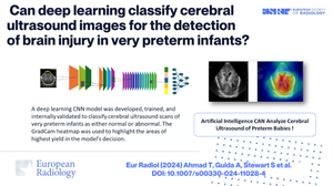
Can deep learning classify cerebral ultrasound images for the detection of brain injury in very preterm infants?
Objectives: Cerebral ultrasound (CUS) is the main imaging screening tool in preterm infants. The aim of this work is to develop deep learning (DL) models that classify normal vs abnormal CUS to serve as a computer-aided detection tool providing timely interpretation of the scans.
Methods: A population-based cohort of very preterm infants (220-306 weeks) born between 2004 and 2016 in Nova Scotia, Canada. A set of nine sequential CUS images per infant was retrieved at three specific coronal landmarks at three pre-identified times (first, sixth weeks, and term age). A radiologist manually labeled each image as normal or abnormal. The dataset was split into training/development/test subsets (80:10:10). Different convolutional neural networks were tested, with filtering of the most uncertain prediction. The model's performance was assessed using precision/recall and the receiver operating area under the curve.
Results: Sequential CUS retrieved for 538/665 babies (81% of the cohort). Four thousand one hundred eighty images were used to develop and test the model. The model performance was only discrete at the beginning but, through different machine learning strategies was boosted to good levels averaging 0.86 ROC AUC (95% CI: 0.82, 0.90) and 0.87 PR AUC (95% CI: 0.84, 0.90) (model uncertainty estimation filters using normalized entropy threshold = 0.5).
Conclusion: This study offers proof of the feasibility of applying DL to CUS. This basic diagnostic model showed good discriminative ability to classify normal versus abnormal CUS. This serves as a CAD and a framework for constructing a prognostic model.
Clinical relevance statement: This DL model can serve as a computer-aided detection tool to classify CUS of very preterm babies as either normal or abnormal. This model will also be used as a framework to develop a prognostic model.
Key points: Binary computer-aided detection models of CUS are applicable for classifying ultrasound images in very preterm babies. This model acts as a step towards developing a model for predicting neurodevelopmental outcomes in very preterm babies. This model serves as a tool for interpretation of CUS in this patient population with a heightened risk of brain injury.
期刊介绍:
European Radiology (ER) continuously updates scientific knowledge in radiology by publication of strong original articles and state-of-the-art reviews written by leading radiologists. A well balanced combination of review articles, original papers, short communications from European radiological congresses and information on society matters makes ER an indispensable source for current information in this field.
This is the Journal of the European Society of Radiology, and the official journal of a number of societies.
From 2004-2008 supplements to European Radiology were published under its companion, European Radiology Supplements, ISSN 1613-3749.

 求助内容:
求助内容: 应助结果提醒方式:


