Tarun Kumar Singh, Ashish J. Johnson, Aakash Gupta, Ikroop Gill
{"title":"利用 CBCT 对宫颈擦伤进行三维分类:综合分析","authors":"Tarun Kumar Singh, Ashish J. Johnson, Aakash Gupta, Ikroop Gill","doi":"10.1016/j.jobcr.2024.08.007","DOIUrl":null,"url":null,"abstract":"<div><h3>Introduction</h3><p>Tooth cervical abrasion (CA) is a prevalent non-carious cervical lesion that poses challenges for accurate diagnosis from periapical radiographs due to difficulties in assessing the lesion's extent, associated bone loss, and pulpal involvement. The presence of overlying bone structures on the palatal side when lesions are located on the buccal side, or vice versa, further complicates radiographic interpretation. So it is important to define the lesions in all three dimensions.</p></div><div><h3>Objective</h3><p>To provide a three-dimensional descriptive classification for cervical abrasion lesions using Cone Beam Computed Tomography (CBCT).</p></div><div><h3>Method</h3><p>A total of 50 patients with cervical abrasion were selected for the study. From these patients, teeth (n = 10) from each of the four different quadrants were chosen. A CBCT scan with a 6 × 6 cm field of view (FOV) was performed, and the DICOM files of the cervical lesions were transferred to 3-D imaging software. The CBCT images of the cervical abrasion lesions were assessed at the level of the deepest point of the lesion along the long axis of the tooth in both axial and sagittal planes. The height (A), buccolingual dimension (B), circumferential spread (C), and remaining dentine thickness (D) were evaluated and classified using new scoring criteria for each dimension. The reliability and reproducibility of the classification were assessed to ensure its clinical applicability.</p></div><div><h3>Conclusion</h3><p>CBCT can be utilized to classify tooth cervical abrasion in endodontics, enhancing diagnosis, analysis, and treatment outcomes. This three-dimensional view facilitates easier communication among clinicians, allows for tailored treatment approaches, and opens new avenues for research.</p></div>","PeriodicalId":16609,"journal":{"name":"Journal of oral biology and craniofacial research","volume":"14 5","pages":"Pages 638-644"},"PeriodicalIF":0.0000,"publicationDate":"2024-08-28","publicationTypes":"Journal Article","fieldsOfStudy":null,"isOpenAccess":false,"openAccessPdf":"https://www.sciencedirect.com/science/article/pii/S2212426824001234/pdfft?md5=494c4c352bdc207748136861f392bcd2&pid=1-s2.0-S2212426824001234-main.pdf","citationCount":"0","resultStr":"{\"title\":\"Novel 3-dimensional classification of cervical abrasion using CBCT: A comprehensive analysis\",\"authors\":\"Tarun Kumar Singh, Ashish J. Johnson, Aakash Gupta, Ikroop Gill\",\"doi\":\"10.1016/j.jobcr.2024.08.007\",\"DOIUrl\":null,\"url\":null,\"abstract\":\"<div><h3>Introduction</h3><p>Tooth cervical abrasion (CA) is a prevalent non-carious cervical lesion that poses challenges for accurate diagnosis from periapical radiographs due to difficulties in assessing the lesion's extent, associated bone loss, and pulpal involvement. The presence of overlying bone structures on the palatal side when lesions are located on the buccal side, or vice versa, further complicates radiographic interpretation. So it is important to define the lesions in all three dimensions.</p></div><div><h3>Objective</h3><p>To provide a three-dimensional descriptive classification for cervical abrasion lesions using Cone Beam Computed Tomography (CBCT).</p></div><div><h3>Method</h3><p>A total of 50 patients with cervical abrasion were selected for the study. From these patients, teeth (n = 10) from each of the four different quadrants were chosen. A CBCT scan with a 6 × 6 cm field of view (FOV) was performed, and the DICOM files of the cervical lesions were transferred to 3-D imaging software. The CBCT images of the cervical abrasion lesions were assessed at the level of the deepest point of the lesion along the long axis of the tooth in both axial and sagittal planes. The height (A), buccolingual dimension (B), circumferential spread (C), and remaining dentine thickness (D) were evaluated and classified using new scoring criteria for each dimension. The reliability and reproducibility of the classification were assessed to ensure its clinical applicability.</p></div><div><h3>Conclusion</h3><p>CBCT can be utilized to classify tooth cervical abrasion in endodontics, enhancing diagnosis, analysis, and treatment outcomes. This three-dimensional view facilitates easier communication among clinicians, allows for tailored treatment approaches, and opens new avenues for research.</p></div>\",\"PeriodicalId\":16609,\"journal\":{\"name\":\"Journal of oral biology and craniofacial research\",\"volume\":\"14 5\",\"pages\":\"Pages 638-644\"},\"PeriodicalIF\":0.0000,\"publicationDate\":\"2024-08-28\",\"publicationTypes\":\"Journal Article\",\"fieldsOfStudy\":null,\"isOpenAccess\":false,\"openAccessPdf\":\"https://www.sciencedirect.com/science/article/pii/S2212426824001234/pdfft?md5=494c4c352bdc207748136861f392bcd2&pid=1-s2.0-S2212426824001234-main.pdf\",\"citationCount\":\"0\",\"resultStr\":null,\"platform\":\"Semanticscholar\",\"paperid\":null,\"PeriodicalName\":\"Journal of oral biology and craniofacial research\",\"FirstCategoryId\":\"1085\",\"ListUrlMain\":\"https://www.sciencedirect.com/science/article/pii/S2212426824001234\",\"RegionNum\":0,\"RegionCategory\":null,\"ArticlePicture\":[],\"TitleCN\":null,\"AbstractTextCN\":null,\"PMCID\":null,\"EPubDate\":\"\",\"PubModel\":\"\",\"JCR\":\"Q1\",\"JCRName\":\"Medicine\",\"Score\":null,\"Total\":0}","platform":"Semanticscholar","paperid":null,"PeriodicalName":"Journal of oral biology and craniofacial research","FirstCategoryId":"1085","ListUrlMain":"https://www.sciencedirect.com/science/article/pii/S2212426824001234","RegionNum":0,"RegionCategory":null,"ArticlePicture":[],"TitleCN":null,"AbstractTextCN":null,"PMCID":null,"EPubDate":"","PubModel":"","JCR":"Q1","JCRName":"Medicine","Score":null,"Total":0}
Novel 3-dimensional classification of cervical abrasion using CBCT: A comprehensive analysis
Introduction
Tooth cervical abrasion (CA) is a prevalent non-carious cervical lesion that poses challenges for accurate diagnosis from periapical radiographs due to difficulties in assessing the lesion's extent, associated bone loss, and pulpal involvement. The presence of overlying bone structures on the palatal side when lesions are located on the buccal side, or vice versa, further complicates radiographic interpretation. So it is important to define the lesions in all three dimensions.
Objective
To provide a three-dimensional descriptive classification for cervical abrasion lesions using Cone Beam Computed Tomography (CBCT).
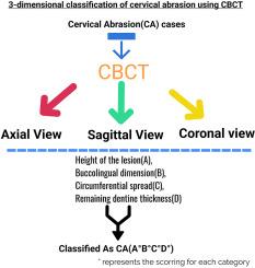
Method
A total of 50 patients with cervical abrasion were selected for the study. From these patients, teeth (n = 10) from each of the four different quadrants were chosen. A CBCT scan with a 6 × 6 cm field of view (FOV) was performed, and the DICOM files of the cervical lesions were transferred to 3-D imaging software. The CBCT images of the cervical abrasion lesions were assessed at the level of the deepest point of the lesion along the long axis of the tooth in both axial and sagittal planes. The height (A), buccolingual dimension (B), circumferential spread (C), and remaining dentine thickness (D) were evaluated and classified using new scoring criteria for each dimension. The reliability and reproducibility of the classification were assessed to ensure its clinical applicability.
Conclusion
CBCT can be utilized to classify tooth cervical abrasion in endodontics, enhancing diagnosis, analysis, and treatment outcomes. This three-dimensional view facilitates easier communication among clinicians, allows for tailored treatment approaches, and opens new avenues for research.
期刊介绍:
Journal of Oral Biology and Craniofacial Research (JOBCR)is the official journal of the Craniofacial Research Foundation (CRF). The journal aims to provide a common platform for both clinical and translational research and to promote interdisciplinary sciences in craniofacial region. JOBCR publishes content that includes diseases, injuries and defects in the head, neck, face, jaws and the hard and soft tissues of the mouth and jaws and face region; diagnosis and medical management of diseases specific to the orofacial tissues and of oral manifestations of systemic diseases; studies on identifying populations at risk of oral disease or in need of specific care, and comparing regional, environmental, social, and access similarities and differences in dental care between populations; diseases of the mouth and related structures like salivary glands, temporomandibular joints, facial muscles and perioral skin; biomedical engineering, tissue engineering and stem cells. The journal publishes reviews, commentaries, peer-reviewed original research articles, short communication, and case reports.

 求助内容:
求助内容: 应助结果提醒方式:


