{"title":"中风诱发的布罗卡失语症的动态程度中心性因第一语言而异:功能性核磁共振成像研究","authors":"Gu Linazi, Sijing Li, Mei Qu, Yanling Xi","doi":"10.1111/jon.13231","DOIUrl":null,"url":null,"abstract":"<div>\n \n \n <section>\n \n <h3> Background and Purpose</h3>\n \n <p>This study sought to explore dynamic degree centrality (DC) variability in particular regions of the brain in patients with poststroke Broca aphasia (BA) using a resting-state functional magnetic resonance imaging (rs-fMRI) approach, comparing differences between Uyghur and Chinese BA patients.</p>\n </section>\n \n <section>\n \n <h3> Methods</h3>\n \n <p>This study investigated two factors, language and BA status, and divided patients into four groups: Uyghur aphasia patients (UA), Uyghur normal control subjects (UN), Chinese aphasia patients (CA), and Chinese normal subjects (CN) who underwent rs-fMRI analysis. Two-way analysis of variance (ANOVA) was used to calculate the comprehensive differences in dynamic DC among these four groups. Correlations between DC and language behavior were assessed with partial correlation analyses.</p>\n </section>\n \n <section>\n \n <h3> Results</h3>\n \n <p>Two-way ANOVA revealed comparable results for the results of pairwise comparisons of dynamic DC variability among the four groups in the right middle frontal gyrus/orbital part (ORBmid.R), right superior frontal gyrus/dorsolateral, and right precuneus (PCUN.R), with results as follows: UA < UN, CA > CN, UA < CA, and UN > CN (<i>p</i> < .05, with the exception of the <i>p</i>-values for UA and UN in superior frontal gyrus/dorsolateral). In contrast, the opposite results were observed for the right calcarine fissure and surrounding cortex (CAL.R, <i>p</i> < .05).</p>\n </section>\n \n <section>\n \n <h3> Conclusion</h3>\n \n <p>The observed enhancement of dynamic DC variability in ORBmid.R and PCUN.R among Chinese BA patients and in CAL.R in Uyghur BA patients may be attributable to language network restructuring. Overall, these results suggest that BA patients who use different language families may exhibit differences in the network mechanisms that characterize observed impairments of language function.</p>\n </section>\n </div>","PeriodicalId":16399,"journal":{"name":"Journal of Neuroimaging","volume":"34 6","pages":"732-741"},"PeriodicalIF":2.3000,"publicationDate":"2024-08-22","publicationTypes":"Journal Article","fieldsOfStudy":null,"isOpenAccess":false,"openAccessPdf":"https://onlinelibrary.wiley.com/doi/epdf/10.1111/jon.13231","citationCount":"0","resultStr":"{\"title\":\"Dynamic degree centrality in stroke-induced Broca's aphasia varies based on first language: A functional MRI study\",\"authors\":\"Gu Linazi, Sijing Li, Mei Qu, Yanling Xi\",\"doi\":\"10.1111/jon.13231\",\"DOIUrl\":null,\"url\":null,\"abstract\":\"<div>\\n \\n \\n <section>\\n \\n <h3> Background and Purpose</h3>\\n \\n <p>This study sought to explore dynamic degree centrality (DC) variability in particular regions of the brain in patients with poststroke Broca aphasia (BA) using a resting-state functional magnetic resonance imaging (rs-fMRI) approach, comparing differences between Uyghur and Chinese BA patients.</p>\\n </section>\\n \\n <section>\\n \\n <h3> Methods</h3>\\n \\n <p>This study investigated two factors, language and BA status, and divided patients into four groups: Uyghur aphasia patients (UA), Uyghur normal control subjects (UN), Chinese aphasia patients (CA), and Chinese normal subjects (CN) who underwent rs-fMRI analysis. Two-way analysis of variance (ANOVA) was used to calculate the comprehensive differences in dynamic DC among these four groups. Correlations between DC and language behavior were assessed with partial correlation analyses.</p>\\n </section>\\n \\n <section>\\n \\n <h3> Results</h3>\\n \\n <p>Two-way ANOVA revealed comparable results for the results of pairwise comparisons of dynamic DC variability among the four groups in the right middle frontal gyrus/orbital part (ORBmid.R), right superior frontal gyrus/dorsolateral, and right precuneus (PCUN.R), with results as follows: UA < UN, CA > CN, UA < CA, and UN > CN (<i>p</i> < .05, with the exception of the <i>p</i>-values for UA and UN in superior frontal gyrus/dorsolateral). In contrast, the opposite results were observed for the right calcarine fissure and surrounding cortex (CAL.R, <i>p</i> < .05).</p>\\n </section>\\n \\n <section>\\n \\n <h3> Conclusion</h3>\\n \\n <p>The observed enhancement of dynamic DC variability in ORBmid.R and PCUN.R among Chinese BA patients and in CAL.R in Uyghur BA patients may be attributable to language network restructuring. Overall, these results suggest that BA patients who use different language families may exhibit differences in the network mechanisms that characterize observed impairments of language function.</p>\\n </section>\\n </div>\",\"PeriodicalId\":16399,\"journal\":{\"name\":\"Journal of Neuroimaging\",\"volume\":\"34 6\",\"pages\":\"732-741\"},\"PeriodicalIF\":2.3000,\"publicationDate\":\"2024-08-22\",\"publicationTypes\":\"Journal Article\",\"fieldsOfStudy\":null,\"isOpenAccess\":false,\"openAccessPdf\":\"https://onlinelibrary.wiley.com/doi/epdf/10.1111/jon.13231\",\"citationCount\":\"0\",\"resultStr\":null,\"platform\":\"Semanticscholar\",\"paperid\":null,\"PeriodicalName\":\"Journal of Neuroimaging\",\"FirstCategoryId\":\"3\",\"ListUrlMain\":\"https://onlinelibrary.wiley.com/doi/10.1111/jon.13231\",\"RegionNum\":4,\"RegionCategory\":\"医学\",\"ArticlePicture\":[],\"TitleCN\":null,\"AbstractTextCN\":null,\"PMCID\":null,\"EPubDate\":\"\",\"PubModel\":\"\",\"JCR\":\"Q3\",\"JCRName\":\"CLINICAL NEUROLOGY\",\"Score\":null,\"Total\":0}","platform":"Semanticscholar","paperid":null,"PeriodicalName":"Journal of Neuroimaging","FirstCategoryId":"3","ListUrlMain":"https://onlinelibrary.wiley.com/doi/10.1111/jon.13231","RegionNum":4,"RegionCategory":"医学","ArticlePicture":[],"TitleCN":null,"AbstractTextCN":null,"PMCID":null,"EPubDate":"","PubModel":"","JCR":"Q3","JCRName":"CLINICAL NEUROLOGY","Score":null,"Total":0}
引用次数: 0
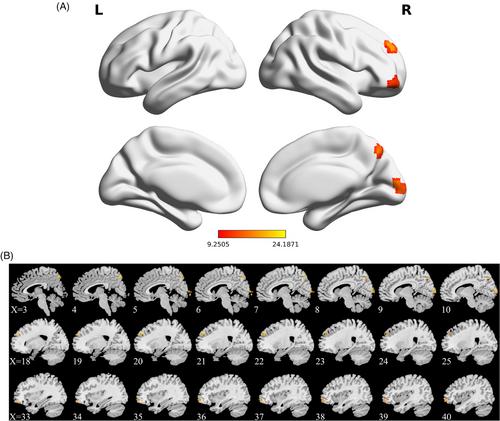
摘要
研究背景与目的:本研究试图利用静息态功能磁共振成像(rs-fMRI)方法探讨脑卒中后布罗卡失语症(BA)患者大脑特定区域的动态度中心性(DC)变异性,比较维吾尔语和汉语BA患者之间的差异:本研究调查了语言和BA状态两个因素,并将患者分为四组:维吾尔语失语症患者(UA)、维吾尔语正常对照组(UN)、汉语失语症患者(CA)和汉语正常对照组(CN)。研究人员采用双向方差分析(ANOVA)计算了四组间动态直流电的综合差异。通过偏相关分析评估了动态直流电与语言行为之间的相关性:双向方差分析显示,四组在右额叶中回/眶部(ORBmid.R)、右额叶上回/背外侧和右楔前(PCUN.R)的动态直流变异性配对比较结果具有可比性,结果如下:UA CN、UA CN(p 结论:在右侧额上回/背外侧和右侧楔前回(PCUN.R)中,观察到动态直流电变异增强:所观察到的汉语 BA 患者 ORBmid.R 和 PCUN.R 以及维吾尔语 BA 患者 CAL.R 的动态直流变异性增强可能归因于语言网络重组。总之,这些结果表明,使用不同语系的 BA 患者可能表现出不同的网络机制,而这些网络机制正是所观察到的语言功能障碍的特征。
Dynamic degree centrality in stroke-induced Broca's aphasia varies based on first language: A functional MRI study
Background and Purpose
This study sought to explore dynamic degree centrality (DC) variability in particular regions of the brain in patients with poststroke Broca aphasia (BA) using a resting-state functional magnetic resonance imaging (rs-fMRI) approach, comparing differences between Uyghur and Chinese BA patients.
Methods
This study investigated two factors, language and BA status, and divided patients into four groups: Uyghur aphasia patients (UA), Uyghur normal control subjects (UN), Chinese aphasia patients (CA), and Chinese normal subjects (CN) who underwent rs-fMRI analysis. Two-way analysis of variance (ANOVA) was used to calculate the comprehensive differences in dynamic DC among these four groups. Correlations between DC and language behavior were assessed with partial correlation analyses.
Results
Two-way ANOVA revealed comparable results for the results of pairwise comparisons of dynamic DC variability among the four groups in the right middle frontal gyrus/orbital part (ORBmid.R), right superior frontal gyrus/dorsolateral, and right precuneus (PCUN.R), with results as follows: UA < UN, CA > CN, UA < CA, and UN > CN (p < .05, with the exception of the p-values for UA and UN in superior frontal gyrus/dorsolateral). In contrast, the opposite results were observed for the right calcarine fissure and surrounding cortex (CAL.R, p < .05).
Conclusion
The observed enhancement of dynamic DC variability in ORBmid.R and PCUN.R among Chinese BA patients and in CAL.R in Uyghur BA patients may be attributable to language network restructuring. Overall, these results suggest that BA patients who use different language families may exhibit differences in the network mechanisms that characterize observed impairments of language function.
期刊介绍:
Start reading the Journal of Neuroimaging to learn the latest neurological imaging techniques. The peer-reviewed research is written in a practical clinical context, giving you the information you need on:
MRI
CT
Carotid Ultrasound and TCD
SPECT
PET
Endovascular Surgical Neuroradiology
Functional MRI
Xenon CT
and other new and upcoming neuroscientific modalities.The Journal of Neuroimaging addresses the full spectrum of human nervous system disease, including stroke, neoplasia, degenerating and demyelinating disease, epilepsy, tumors, lesions, infectious disease, cerebral vascular arterial diseases, toxic-metabolic disease, psychoses, dementias, heredo-familial disease, and trauma.Offering original research, review articles, case reports, neuroimaging CPCs, and evaluations of instruments and technology relevant to the nervous system, the Journal of Neuroimaging focuses on useful clinical developments and applications, tested techniques and interpretations, patient care, diagnostics, and therapeutics. Start reading today!

 求助内容:
求助内容: 应助结果提醒方式:


