Louis Gagnon, Diviya Gupta, George Mastorakos, Nathan White, Vanessa Goodwill, Carrie R McDonald, Thomas Beaumont, Christopher Conlin, Tyler M Seibert, Uyen Nguyen, Jona Hattangadi-Gluth, Santosh Kesari, Jessica D Schulte, David Piccioni, Kathleen M Schmainda, Nikdokht Farid, Anders M Dale, Jeffrey D Rudie
下载PDF
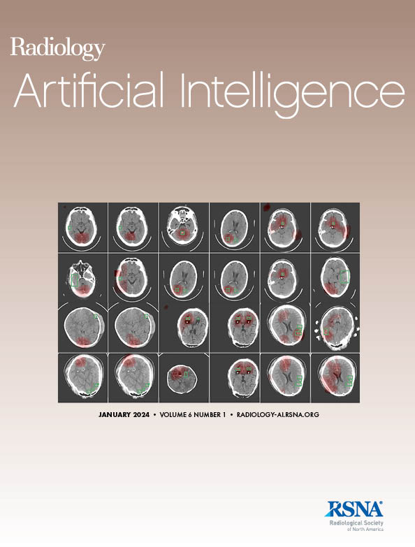
{"title":"胶质母细胞瘤治疗前和治疗后多壳体弥散 MRI 上浸润性和增强型细胞肿瘤的深度学习分割","authors":"Louis Gagnon, Diviya Gupta, George Mastorakos, Nathan White, Vanessa Goodwill, Carrie R McDonald, Thomas Beaumont, Christopher Conlin, Tyler M Seibert, Uyen Nguyen, Jona Hattangadi-Gluth, Santosh Kesari, Jessica D Schulte, David Piccioni, Kathleen M Schmainda, Nikdokht Farid, Anders M Dale, Jeffrey D Rudie","doi":"10.1148/ryai.230489","DOIUrl":null,"url":null,"abstract":"<p><p>Purpose To develop and validate a deep learning (DL) method to detect and segment enhancing and nonenhancing cellular tumor on pre- and posttreatment MRI scans in patients with glioblastoma and to predict overall survival (OS) and progression-free survival (PFS). Materials and Methods This retrospective study included 1397 MRI scans in 1297 patients with glioblastoma, including an internal set of 243 MRI scans (January 2010 to June 2022) for model training and cross-validation and four external test cohorts. Cellular tumor maps were segmented by two radiologists on the basis of imaging, clinical history, and pathologic findings. Multimodal MRI data with perfusion and multishell diffusion imaging were inputted into a nnU-Net DL model to segment cellular tumor. Segmentation performance (Dice score) and performance in distinguishing recurrent tumor from posttreatment changes (area under the receiver operating characteristic curve [AUC]) were quantified. Model performance in predicting OS and PFS was assessed using Cox multivariable analysis. Results A cohort of 178 patients (mean age, 56 years ± 13 [SD]; 116 male, 62 female) with 243 MRI timepoints, as well as four external datasets with 55, 70, 610, and 419 MRI timepoints, respectively, were evaluated. The median Dice score was 0.79 (IQR, 0.53-0.89), and the AUC for detecting residual or recurrent tumor was 0.84 (95% CI: 0.79, 0.89). In the internal test set, estimated cellular tumor volume was significantly associated with OS (hazard ratio [HR] = 1.04 per milliliter; <i>P</i> < .001) and PFS (HR = 1.04 per milliliter; <i>P</i> < .001) after adjustment for age, sex, and gross total resection (GTR) status. In the external test sets, estimated cellular tumor volume was significantly associated with OS (HR = 1.01 per milliliter; <i>P</i> < .001) after adjustment for age, sex, and GTR status. Conclusion A DL model incorporating advanced imaging could accurately segment enhancing and nonenhancing cellular tumor, distinguish recurrent or residual tumor from posttreatment changes, and predict OS and PFS in patients with glioblastoma. <b>Keywords:</b> Segmentation, Glioblastoma, Multishell Diffusion MRI <i>Supplemental material is available for this article.</i> © RSNA, 2024.</p>","PeriodicalId":29787,"journal":{"name":"Radiology-Artificial Intelligence","volume":" ","pages":"e230489"},"PeriodicalIF":8.1000,"publicationDate":"2024-09-01","publicationTypes":"Journal Article","fieldsOfStudy":null,"isOpenAccess":false,"openAccessPdf":"https://www.ncbi.nlm.nih.gov/pmc/articles/PMC11427928/pdf/","citationCount":"0","resultStr":"{\"title\":\"Deep Learning Segmentation of Infiltrative and Enhancing Cellular Tumor at Pre- and Posttreatment Multishell Diffusion MRI of Glioblastoma.\",\"authors\":\"Louis Gagnon, Diviya Gupta, George Mastorakos, Nathan White, Vanessa Goodwill, Carrie R McDonald, Thomas Beaumont, Christopher Conlin, Tyler M Seibert, Uyen Nguyen, Jona Hattangadi-Gluth, Santosh Kesari, Jessica D Schulte, David Piccioni, Kathleen M Schmainda, Nikdokht Farid, Anders M Dale, Jeffrey D Rudie\",\"doi\":\"10.1148/ryai.230489\",\"DOIUrl\":null,\"url\":null,\"abstract\":\"<p><p>Purpose To develop and validate a deep learning (DL) method to detect and segment enhancing and nonenhancing cellular tumor on pre- and posttreatment MRI scans in patients with glioblastoma and to predict overall survival (OS) and progression-free survival (PFS). Materials and Methods This retrospective study included 1397 MRI scans in 1297 patients with glioblastoma, including an internal set of 243 MRI scans (January 2010 to June 2022) for model training and cross-validation and four external test cohorts. Cellular tumor maps were segmented by two radiologists on the basis of imaging, clinical history, and pathologic findings. Multimodal MRI data with perfusion and multishell diffusion imaging were inputted into a nnU-Net DL model to segment cellular tumor. Segmentation performance (Dice score) and performance in distinguishing recurrent tumor from posttreatment changes (area under the receiver operating characteristic curve [AUC]) were quantified. Model performance in predicting OS and PFS was assessed using Cox multivariable analysis. Results A cohort of 178 patients (mean age, 56 years ± 13 [SD]; 116 male, 62 female) with 243 MRI timepoints, as well as four external datasets with 55, 70, 610, and 419 MRI timepoints, respectively, were evaluated. The median Dice score was 0.79 (IQR, 0.53-0.89), and the AUC for detecting residual or recurrent tumor was 0.84 (95% CI: 0.79, 0.89). In the internal test set, estimated cellular tumor volume was significantly associated with OS (hazard ratio [HR] = 1.04 per milliliter; <i>P</i> < .001) and PFS (HR = 1.04 per milliliter; <i>P</i> < .001) after adjustment for age, sex, and gross total resection (GTR) status. In the external test sets, estimated cellular tumor volume was significantly associated with OS (HR = 1.01 per milliliter; <i>P</i> < .001) after adjustment for age, sex, and GTR status. Conclusion A DL model incorporating advanced imaging could accurately segment enhancing and nonenhancing cellular tumor, distinguish recurrent or residual tumor from posttreatment changes, and predict OS and PFS in patients with glioblastoma. <b>Keywords:</b> Segmentation, Glioblastoma, Multishell Diffusion MRI <i>Supplemental material is available for this article.</i> © RSNA, 2024.</p>\",\"PeriodicalId\":29787,\"journal\":{\"name\":\"Radiology-Artificial Intelligence\",\"volume\":\" \",\"pages\":\"e230489\"},\"PeriodicalIF\":8.1000,\"publicationDate\":\"2024-09-01\",\"publicationTypes\":\"Journal Article\",\"fieldsOfStudy\":null,\"isOpenAccess\":false,\"openAccessPdf\":\"https://www.ncbi.nlm.nih.gov/pmc/articles/PMC11427928/pdf/\",\"citationCount\":\"0\",\"resultStr\":null,\"platform\":\"Semanticscholar\",\"paperid\":null,\"PeriodicalName\":\"Radiology-Artificial Intelligence\",\"FirstCategoryId\":\"1085\",\"ListUrlMain\":\"https://doi.org/10.1148/ryai.230489\",\"RegionNum\":0,\"RegionCategory\":null,\"ArticlePicture\":[],\"TitleCN\":null,\"AbstractTextCN\":null,\"PMCID\":null,\"EPubDate\":\"\",\"PubModel\":\"\",\"JCR\":\"Q1\",\"JCRName\":\"COMPUTER SCIENCE, ARTIFICIAL INTELLIGENCE\",\"Score\":null,\"Total\":0}","platform":"Semanticscholar","paperid":null,"PeriodicalName":"Radiology-Artificial Intelligence","FirstCategoryId":"1085","ListUrlMain":"https://doi.org/10.1148/ryai.230489","RegionNum":0,"RegionCategory":null,"ArticlePicture":[],"TitleCN":null,"AbstractTextCN":null,"PMCID":null,"EPubDate":"","PubModel":"","JCR":"Q1","JCRName":"COMPUTER SCIENCE, ARTIFICIAL INTELLIGENCE","Score":null,"Total":0}
引用次数: 0
引用
批量引用

 求助内容:
求助内容: 应助结果提醒方式:


