Sandra Schuh, Maximilian Berger, Stefan Schiele, Anna Rubeck, Gernot Müller, Jennifer Jahel Vélez González, Jon Holmes, Julia Welzel
{"title":"用于急性伤口愈合成像的动态光学相干断层扫描。","authors":"Sandra Schuh, Maximilian Berger, Stefan Schiele, Anna Rubeck, Gernot Müller, Jennifer Jahel Vélez González, Jon Holmes, Julia Welzel","doi":"10.1111/iwj.70015","DOIUrl":null,"url":null,"abstract":"<p>The aim of this study was to investigate acute wound healing with dynamic optical coherence tomography (D-OCT). From 22 patients with 23 split skin graft donor sites, vessels at four wound edges, the wound bed, and adjacent and unaffected skin of the contralateral leg were measured by D-OCT at six time points from surgery to 4 weeks of healing. Changes in vessel orientation, density, diameter, morphology and pattern in horizontal, vertical and 3D images were analysed for wound healing and re-epithelialization. At 300 μm depth, there were significant differences of blobs and serpiginous vessels between normal and wounded skin. The wound had significantly more vertically oriented vessels, a higher degree of branching, vessel density and diameter compared with healthy skin. 3D images showed increased angiogenesis from healthy skin towards the wound centre, significantly higher vessel density at the wound than at normal skin and the highest at the interface. During wound healing blobs, coils and serpiginous vessels occurred significantly more frequently in lesional than healthy skin. Vessel density was greatest at the beginning, decreased and then increased by 4 weeks post-surgery. D-OCT helps to evaluate acute wound healing by visualizing and quantifying blood vessel growth in addition to re-epithelialization.</p>","PeriodicalId":14451,"journal":{"name":"International Wound Journal","volume":null,"pages":null},"PeriodicalIF":2.6000,"publicationDate":"2024-08-20","publicationTypes":"Journal Article","fieldsOfStudy":null,"isOpenAccess":false,"openAccessPdf":"https://onlinelibrary.wiley.com/doi/epdf/10.1111/iwj.70015","citationCount":"0","resultStr":"{\"title\":\"Dynamic optical coherence tomography for imaging acute wound healing\",\"authors\":\"Sandra Schuh, Maximilian Berger, Stefan Schiele, Anna Rubeck, Gernot Müller, Jennifer Jahel Vélez González, Jon Holmes, Julia Welzel\",\"doi\":\"10.1111/iwj.70015\",\"DOIUrl\":null,\"url\":null,\"abstract\":\"<p>The aim of this study was to investigate acute wound healing with dynamic optical coherence tomography (D-OCT). From 22 patients with 23 split skin graft donor sites, vessels at four wound edges, the wound bed, and adjacent and unaffected skin of the contralateral leg were measured by D-OCT at six time points from surgery to 4 weeks of healing. Changes in vessel orientation, density, diameter, morphology and pattern in horizontal, vertical and 3D images were analysed for wound healing and re-epithelialization. At 300 μm depth, there were significant differences of blobs and serpiginous vessels between normal and wounded skin. The wound had significantly more vertically oriented vessels, a higher degree of branching, vessel density and diameter compared with healthy skin. 3D images showed increased angiogenesis from healthy skin towards the wound centre, significantly higher vessel density at the wound than at normal skin and the highest at the interface. During wound healing blobs, coils and serpiginous vessels occurred significantly more frequently in lesional than healthy skin. Vessel density was greatest at the beginning, decreased and then increased by 4 weeks post-surgery. D-OCT helps to evaluate acute wound healing by visualizing and quantifying blood vessel growth in addition to re-epithelialization.</p>\",\"PeriodicalId\":14451,\"journal\":{\"name\":\"International Wound Journal\",\"volume\":null,\"pages\":null},\"PeriodicalIF\":2.6000,\"publicationDate\":\"2024-08-20\",\"publicationTypes\":\"Journal Article\",\"fieldsOfStudy\":null,\"isOpenAccess\":false,\"openAccessPdf\":\"https://onlinelibrary.wiley.com/doi/epdf/10.1111/iwj.70015\",\"citationCount\":\"0\",\"resultStr\":null,\"platform\":\"Semanticscholar\",\"paperid\":null,\"PeriodicalName\":\"International Wound Journal\",\"FirstCategoryId\":\"3\",\"ListUrlMain\":\"https://onlinelibrary.wiley.com/doi/10.1111/iwj.70015\",\"RegionNum\":3,\"RegionCategory\":\"医学\",\"ArticlePicture\":[],\"TitleCN\":null,\"AbstractTextCN\":null,\"PMCID\":null,\"EPubDate\":\"\",\"PubModel\":\"\",\"JCR\":\"Q2\",\"JCRName\":\"DERMATOLOGY\",\"Score\":null,\"Total\":0}","platform":"Semanticscholar","paperid":null,"PeriodicalName":"International Wound Journal","FirstCategoryId":"3","ListUrlMain":"https://onlinelibrary.wiley.com/doi/10.1111/iwj.70015","RegionNum":3,"RegionCategory":"医学","ArticlePicture":[],"TitleCN":null,"AbstractTextCN":null,"PMCID":null,"EPubDate":"","PubModel":"","JCR":"Q2","JCRName":"DERMATOLOGY","Score":null,"Total":0}
Dynamic optical coherence tomography for imaging acute wound healing
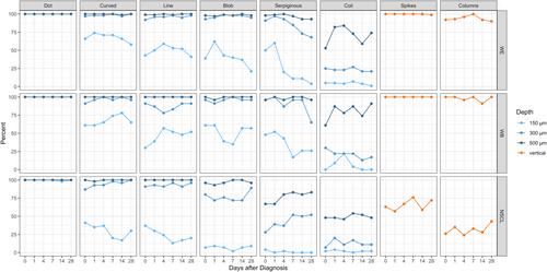
The aim of this study was to investigate acute wound healing with dynamic optical coherence tomography (D-OCT). From 22 patients with 23 split skin graft donor sites, vessels at four wound edges, the wound bed, and adjacent and unaffected skin of the contralateral leg were measured by D-OCT at six time points from surgery to 4 weeks of healing. Changes in vessel orientation, density, diameter, morphology and pattern in horizontal, vertical and 3D images were analysed for wound healing and re-epithelialization. At 300 μm depth, there were significant differences of blobs and serpiginous vessels between normal and wounded skin. The wound had significantly more vertically oriented vessels, a higher degree of branching, vessel density and diameter compared with healthy skin. 3D images showed increased angiogenesis from healthy skin towards the wound centre, significantly higher vessel density at the wound than at normal skin and the highest at the interface. During wound healing blobs, coils and serpiginous vessels occurred significantly more frequently in lesional than healthy skin. Vessel density was greatest at the beginning, decreased and then increased by 4 weeks post-surgery. D-OCT helps to evaluate acute wound healing by visualizing and quantifying blood vessel growth in addition to re-epithelialization.
期刊介绍:
The Editors welcome papers on all aspects of prevention and treatment of wounds and associated conditions in the fields of surgery, dermatology, oncology, nursing, radiotherapy, physical therapy, occupational therapy and podiatry. The Journal accepts papers in the following categories:
- Research papers
- Review articles
- Clinical studies
- Letters
- News and Views: international perspectives, education initiatives, guidelines and different activities of groups and societies.
Calendar of events
The Editors are supported by a board of international experts and a panel of reviewers across a range of disciplines and specialties which ensures only the most current and relevant research is published.
 求助内容:
求助内容: 应助结果提醒方式:


