Sebastian Ziegelmayer, Alexander W Marka, Maximilian Strenzke, Tristan Lemke, Hannah Rosenkranz, Bernadette Scherer, Thomas Huber, Kilian Weiss, Marcus R Makowski, Dimitrios C Karampinos, Markus Graf, Joshua Gawlitza
{"title":"速度与效率:评估人工智能增强三维梯度回波成像的肺结节检测。","authors":"Sebastian Ziegelmayer, Alexander W Marka, Maximilian Strenzke, Tristan Lemke, Hannah Rosenkranz, Bernadette Scherer, Thomas Huber, Kilian Weiss, Marcus R Makowski, Dimitrios C Karampinos, Markus Graf, Joshua Gawlitza","doi":"10.1007/s00330-024-11027-5","DOIUrl":null,"url":null,"abstract":"<p><strong>Objectives: </strong>Evaluating the diagnostic feasibility of accelerated pulmonary MR imaging for detection and characterisation of pulmonary nodules with artificial intelligence-aided compressed sensing.</p><p><strong>Materials and methods: </strong>In this prospective trial, patients with benign and malignant lung nodules admitted between December 2021 and December 2022 underwent chest CT and pulmonary MRI. Pulmonary MRI used a respiratory-gated 3D gradient echo sequence, accelerated with a combination of parallel imaging, compressed sensing, and deep learning image reconstruction with three different acceleration factors (CS-AI-7, CS-AI-10, and CS-AI-15). Two readers evaluated image quality (5-point Likert scale), nodule detection and characterisation (size and morphology) of all sequences compared to CT in a blinded setting. Reader agreement was determined using the intraclass correlation coefficient (ICC).</p><p><strong>Results: </strong>Thirty-seven patients with 64 pulmonary nodules (solid n = 57 [3-107 mm] part-solid n = 6 [ground glass/solid 8 mm/4-28 mm/16 mm] ground glass nodule n = 1 [20 mm]) were analysed. Nominal scan times were CS-AI-7 3:53 min; CS-AI-10 2:34 min; CS-AI-15 1:50 min. CS-AI-7 showed higher image quality, while quality remained diagnostic even for CS-AI-15. Detection rates of pulmonary nodules were 100%, 98.4%, and 96.8% for CS-AI factors 7, 10, and 15, respectively. Nodule morphology was best at the lowest acceleration and was inferior to CT in only 5% of cases, compared to 10% for CS-AI-10 and 23% for CS-AI-15. The nodule size was comparable for all sequences and deviated on average < 1 mm from the CT size.</p><p><strong>Conclusion: </strong>The combination of compressed sensing and AI enables a substantial reduction in the scan time of lung MRI while maintaining a high detection rate of pulmonary nodules.</p><p><strong>Clinical relevance statement: </strong>Incorporating compressed sensing and AI in pulmonary MRI achieves significant time savings without compromising nodule detection or characteristics. This advancement holds clinical promise, enhancing efficiency in lung cancer screening without sacrificing diagnostic quality.</p><p><strong>Key points: </strong>Lung cancer screening by MRI may be possible but would benefit from scan time optimisation. Significant scan time reduction, high detection rates, and preserved nodule characteristics were achieved across different acceleration factors. Integrating compressed sensing and AI in pulmonary MRI offers efficient lung cancer screening without compromising diagnostic quality.</p>","PeriodicalId":12076,"journal":{"name":"European Radiology","volume":" ","pages":"2237-2244"},"PeriodicalIF":4.7000,"publicationDate":"2025-04-01","publicationTypes":"Journal Article","fieldsOfStudy":null,"isOpenAccess":false,"openAccessPdf":"https://www.ncbi.nlm.nih.gov/pmc/articles/PMC11914225/pdf/","citationCount":"0","resultStr":"{\"title\":\"Speed and efficiency: evaluating pulmonary nodule detection with AI-enhanced 3D gradient echo imaging.\",\"authors\":\"Sebastian Ziegelmayer, Alexander W Marka, Maximilian Strenzke, Tristan Lemke, Hannah Rosenkranz, Bernadette Scherer, Thomas Huber, Kilian Weiss, Marcus R Makowski, Dimitrios C Karampinos, Markus Graf, Joshua Gawlitza\",\"doi\":\"10.1007/s00330-024-11027-5\",\"DOIUrl\":null,\"url\":null,\"abstract\":\"<p><strong>Objectives: </strong>Evaluating the diagnostic feasibility of accelerated pulmonary MR imaging for detection and characterisation of pulmonary nodules with artificial intelligence-aided compressed sensing.</p><p><strong>Materials and methods: </strong>In this prospective trial, patients with benign and malignant lung nodules admitted between December 2021 and December 2022 underwent chest CT and pulmonary MRI. Pulmonary MRI used a respiratory-gated 3D gradient echo sequence, accelerated with a combination of parallel imaging, compressed sensing, and deep learning image reconstruction with three different acceleration factors (CS-AI-7, CS-AI-10, and CS-AI-15). Two readers evaluated image quality (5-point Likert scale), nodule detection and characterisation (size and morphology) of all sequences compared to CT in a blinded setting. Reader agreement was determined using the intraclass correlation coefficient (ICC).</p><p><strong>Results: </strong>Thirty-seven patients with 64 pulmonary nodules (solid n = 57 [3-107 mm] part-solid n = 6 [ground glass/solid 8 mm/4-28 mm/16 mm] ground glass nodule n = 1 [20 mm]) were analysed. Nominal scan times were CS-AI-7 3:53 min; CS-AI-10 2:34 min; CS-AI-15 1:50 min. CS-AI-7 showed higher image quality, while quality remained diagnostic even for CS-AI-15. Detection rates of pulmonary nodules were 100%, 98.4%, and 96.8% for CS-AI factors 7, 10, and 15, respectively. Nodule morphology was best at the lowest acceleration and was inferior to CT in only 5% of cases, compared to 10% for CS-AI-10 and 23% for CS-AI-15. The nodule size was comparable for all sequences and deviated on average < 1 mm from the CT size.</p><p><strong>Conclusion: </strong>The combination of compressed sensing and AI enables a substantial reduction in the scan time of lung MRI while maintaining a high detection rate of pulmonary nodules.</p><p><strong>Clinical relevance statement: </strong>Incorporating compressed sensing and AI in pulmonary MRI achieves significant time savings without compromising nodule detection or characteristics. This advancement holds clinical promise, enhancing efficiency in lung cancer screening without sacrificing diagnostic quality.</p><p><strong>Key points: </strong>Lung cancer screening by MRI may be possible but would benefit from scan time optimisation. Significant scan time reduction, high detection rates, and preserved nodule characteristics were achieved across different acceleration factors. Integrating compressed sensing and AI in pulmonary MRI offers efficient lung cancer screening without compromising diagnostic quality.</p>\",\"PeriodicalId\":12076,\"journal\":{\"name\":\"European Radiology\",\"volume\":\" \",\"pages\":\"2237-2244\"},\"PeriodicalIF\":4.7000,\"publicationDate\":\"2025-04-01\",\"publicationTypes\":\"Journal Article\",\"fieldsOfStudy\":null,\"isOpenAccess\":false,\"openAccessPdf\":\"https://www.ncbi.nlm.nih.gov/pmc/articles/PMC11914225/pdf/\",\"citationCount\":\"0\",\"resultStr\":null,\"platform\":\"Semanticscholar\",\"paperid\":null,\"PeriodicalName\":\"European Radiology\",\"FirstCategoryId\":\"3\",\"ListUrlMain\":\"https://doi.org/10.1007/s00330-024-11027-5\",\"RegionNum\":2,\"RegionCategory\":\"医学\",\"ArticlePicture\":[],\"TitleCN\":null,\"AbstractTextCN\":null,\"PMCID\":null,\"EPubDate\":\"2024/8/18 0:00:00\",\"PubModel\":\"Epub\",\"JCR\":\"Q1\",\"JCRName\":\"RADIOLOGY, NUCLEAR MEDICINE & MEDICAL IMAGING\",\"Score\":null,\"Total\":0}","platform":"Semanticscholar","paperid":null,"PeriodicalName":"European Radiology","FirstCategoryId":"3","ListUrlMain":"https://doi.org/10.1007/s00330-024-11027-5","RegionNum":2,"RegionCategory":"医学","ArticlePicture":[],"TitleCN":null,"AbstractTextCN":null,"PMCID":null,"EPubDate":"2024/8/18 0:00:00","PubModel":"Epub","JCR":"Q1","JCRName":"RADIOLOGY, NUCLEAR MEDICINE & MEDICAL IMAGING","Score":null,"Total":0}
Speed and efficiency: evaluating pulmonary nodule detection with AI-enhanced 3D gradient echo imaging.
Objectives: Evaluating the diagnostic feasibility of accelerated pulmonary MR imaging for detection and characterisation of pulmonary nodules with artificial intelligence-aided compressed sensing.
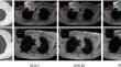
Materials and methods: In this prospective trial, patients with benign and malignant lung nodules admitted between December 2021 and December 2022 underwent chest CT and pulmonary MRI. Pulmonary MRI used a respiratory-gated 3D gradient echo sequence, accelerated with a combination of parallel imaging, compressed sensing, and deep learning image reconstruction with three different acceleration factors (CS-AI-7, CS-AI-10, and CS-AI-15). Two readers evaluated image quality (5-point Likert scale), nodule detection and characterisation (size and morphology) of all sequences compared to CT in a blinded setting. Reader agreement was determined using the intraclass correlation coefficient (ICC).
Results: Thirty-seven patients with 64 pulmonary nodules (solid n = 57 [3-107 mm] part-solid n = 6 [ground glass/solid 8 mm/4-28 mm/16 mm] ground glass nodule n = 1 [20 mm]) were analysed. Nominal scan times were CS-AI-7 3:53 min; CS-AI-10 2:34 min; CS-AI-15 1:50 min. CS-AI-7 showed higher image quality, while quality remained diagnostic even for CS-AI-15. Detection rates of pulmonary nodules were 100%, 98.4%, and 96.8% for CS-AI factors 7, 10, and 15, respectively. Nodule morphology was best at the lowest acceleration and was inferior to CT in only 5% of cases, compared to 10% for CS-AI-10 and 23% for CS-AI-15. The nodule size was comparable for all sequences and deviated on average < 1 mm from the CT size.
Conclusion: The combination of compressed sensing and AI enables a substantial reduction in the scan time of lung MRI while maintaining a high detection rate of pulmonary nodules.
Clinical relevance statement: Incorporating compressed sensing and AI in pulmonary MRI achieves significant time savings without compromising nodule detection or characteristics. This advancement holds clinical promise, enhancing efficiency in lung cancer screening without sacrificing diagnostic quality.
Key points: Lung cancer screening by MRI may be possible but would benefit from scan time optimisation. Significant scan time reduction, high detection rates, and preserved nodule characteristics were achieved across different acceleration factors. Integrating compressed sensing and AI in pulmonary MRI offers efficient lung cancer screening without compromising diagnostic quality.
期刊介绍:
European Radiology (ER) continuously updates scientific knowledge in radiology by publication of strong original articles and state-of-the-art reviews written by leading radiologists. A well balanced combination of review articles, original papers, short communications from European radiological congresses and information on society matters makes ER an indispensable source for current information in this field.
This is the Journal of the European Society of Radiology, and the official journal of a number of societies.
From 2004-2008 supplements to European Radiology were published under its companion, European Radiology Supplements, ISSN 1613-3749.

 求助内容:
求助内容: 应助结果提醒方式:


