{"title":"散发性肌萎缩性脊髓侧索硬化症脊髓运动神经元变性的形态计量分析。","authors":"","doi":"10.1016/j.jns.2024.123177","DOIUrl":null,"url":null,"abstract":"<div><h3>Objectives</h3><p>This study aimed to clarify the relationship between 43-kDa TAR DNA-binding protein (TDP-43) pathology and spinal cord anterior horn motor neuron (AHMN) atrophy in sporadic amyotrophic lateral sclerosis (SALS).</p></div><div><h3>Methods</h3><p>Eight patients with SALS and 12 controls were included in this study. Formalin-fixed specimens of lumbar spinal cord samples were paraffin-embedded and sectioned at the level of the fourth lumbar spinal cord with a 4 μm thickness. Using a microscope, the long diameters of the neurons with nucleoli were measured in spinal AHMNs stained with an anti-SMI-32 antibody. AHMNs were divided into medial and lateral nuclei for statistical analysis. We also used previously reported data to measure the long diameter of AHMNs with initial TDP-43 pathology, in which TDP-43 was present both in the nucleus and cytoplasm.</p></div><div><h3>Results</h3><p>The long diameter of the lumbar spinal AHMNs in patients with SALS was smaller in the medial nucleus (42.54 ± 9.33 μm, <em>n</em> = 24) and the lateral nucleus (49.41 ± 13.86 μm, <em>n</em> = 129) than in controls (medial nucleus: 55.84 ± 13.49 μm, <em>n</em> = 85, <em>p</em> < 0.001; lateral nucleus: 62.39 ± 13.29 μm, <em>n</em> = 756, p < 0.001, Mann–Whitney <em>U</em> test). All 21 motor neurons with initial TDP-43 pathology were in the lateral nucleus, and their long diameter (67.60 ± 18.3 μm, <em>p</em> = 0.352) was not significantly different from that of controls.</p></div><div><h3>Conclusion</h3><p>Motor neuron atrophy in SALS does not occur during the initial stages of TDP-43 pathology, and TDP-43 pathology is already advanced in the atrophied motor neurons.</p></div>","PeriodicalId":17417,"journal":{"name":"Journal of the Neurological Sciences","volume":null,"pages":null},"PeriodicalIF":3.6000,"publicationDate":"2024-08-12","publicationTypes":"Journal Article","fieldsOfStudy":null,"isOpenAccess":false,"openAccessPdf":"","citationCount":"0","resultStr":"{\"title\":\"Morphometric analysis of spinal motor neuron degeneration in sporadic amyotrophic lateral sclerosis\",\"authors\":\"\",\"doi\":\"10.1016/j.jns.2024.123177\",\"DOIUrl\":null,\"url\":null,\"abstract\":\"<div><h3>Objectives</h3><p>This study aimed to clarify the relationship between 43-kDa TAR DNA-binding protein (TDP-43) pathology and spinal cord anterior horn motor neuron (AHMN) atrophy in sporadic amyotrophic lateral sclerosis (SALS).</p></div><div><h3>Methods</h3><p>Eight patients with SALS and 12 controls were included in this study. Formalin-fixed specimens of lumbar spinal cord samples were paraffin-embedded and sectioned at the level of the fourth lumbar spinal cord with a 4 μm thickness. Using a microscope, the long diameters of the neurons with nucleoli were measured in spinal AHMNs stained with an anti-SMI-32 antibody. AHMNs were divided into medial and lateral nuclei for statistical analysis. We also used previously reported data to measure the long diameter of AHMNs with initial TDP-43 pathology, in which TDP-43 was present both in the nucleus and cytoplasm.</p></div><div><h3>Results</h3><p>The long diameter of the lumbar spinal AHMNs in patients with SALS was smaller in the medial nucleus (42.54 ± 9.33 μm, <em>n</em> = 24) and the lateral nucleus (49.41 ± 13.86 μm, <em>n</em> = 129) than in controls (medial nucleus: 55.84 ± 13.49 μm, <em>n</em> = 85, <em>p</em> < 0.001; lateral nucleus: 62.39 ± 13.29 μm, <em>n</em> = 756, p < 0.001, Mann–Whitney <em>U</em> test). All 21 motor neurons with initial TDP-43 pathology were in the lateral nucleus, and their long diameter (67.60 ± 18.3 μm, <em>p</em> = 0.352) was not significantly different from that of controls.</p></div><div><h3>Conclusion</h3><p>Motor neuron atrophy in SALS does not occur during the initial stages of TDP-43 pathology, and TDP-43 pathology is already advanced in the atrophied motor neurons.</p></div>\",\"PeriodicalId\":17417,\"journal\":{\"name\":\"Journal of the Neurological Sciences\",\"volume\":null,\"pages\":null},\"PeriodicalIF\":3.6000,\"publicationDate\":\"2024-08-12\",\"publicationTypes\":\"Journal Article\",\"fieldsOfStudy\":null,\"isOpenAccess\":false,\"openAccessPdf\":\"\",\"citationCount\":\"0\",\"resultStr\":null,\"platform\":\"Semanticscholar\",\"paperid\":null,\"PeriodicalName\":\"Journal of the Neurological Sciences\",\"FirstCategoryId\":\"3\",\"ListUrlMain\":\"https://www.sciencedirect.com/science/article/pii/S0022510X24003125\",\"RegionNum\":3,\"RegionCategory\":\"医学\",\"ArticlePicture\":[],\"TitleCN\":null,\"AbstractTextCN\":null,\"PMCID\":null,\"EPubDate\":\"\",\"PubModel\":\"\",\"JCR\":\"Q1\",\"JCRName\":\"CLINICAL NEUROLOGY\",\"Score\":null,\"Total\":0}","platform":"Semanticscholar","paperid":null,"PeriodicalName":"Journal of the Neurological Sciences","FirstCategoryId":"3","ListUrlMain":"https://www.sciencedirect.com/science/article/pii/S0022510X24003125","RegionNum":3,"RegionCategory":"医学","ArticlePicture":[],"TitleCN":null,"AbstractTextCN":null,"PMCID":null,"EPubDate":"","PubModel":"","JCR":"Q1","JCRName":"CLINICAL NEUROLOGY","Score":null,"Total":0}
Morphometric analysis of spinal motor neuron degeneration in sporadic amyotrophic lateral sclerosis
Objectives
This study aimed to clarify the relationship between 43-kDa TAR DNA-binding protein (TDP-43) pathology and spinal cord anterior horn motor neuron (AHMN) atrophy in sporadic amyotrophic lateral sclerosis (SALS).
Methods
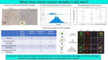
Eight patients with SALS and 12 controls were included in this study. Formalin-fixed specimens of lumbar spinal cord samples were paraffin-embedded and sectioned at the level of the fourth lumbar spinal cord with a 4 μm thickness. Using a microscope, the long diameters of the neurons with nucleoli were measured in spinal AHMNs stained with an anti-SMI-32 antibody. AHMNs were divided into medial and lateral nuclei for statistical analysis. We also used previously reported data to measure the long diameter of AHMNs with initial TDP-43 pathology, in which TDP-43 was present both in the nucleus and cytoplasm.
Results
The long diameter of the lumbar spinal AHMNs in patients with SALS was smaller in the medial nucleus (42.54 ± 9.33 μm, n = 24) and the lateral nucleus (49.41 ± 13.86 μm, n = 129) than in controls (medial nucleus: 55.84 ± 13.49 μm, n = 85, p < 0.001; lateral nucleus: 62.39 ± 13.29 μm, n = 756, p < 0.001, Mann–Whitney U test). All 21 motor neurons with initial TDP-43 pathology were in the lateral nucleus, and their long diameter (67.60 ± 18.3 μm, p = 0.352) was not significantly different from that of controls.
Conclusion
Motor neuron atrophy in SALS does not occur during the initial stages of TDP-43 pathology, and TDP-43 pathology is already advanced in the atrophied motor neurons.
期刊介绍:
The Journal of the Neurological Sciences provides a medium for the prompt publication of original articles in neurology and neuroscience from around the world. JNS places special emphasis on articles that: 1) provide guidance to clinicians around the world (Best Practices, Global Neurology); 2) report cutting-edge science related to neurology (Basic and Translational Sciences); 3) educate readers about relevant and practical clinical outcomes in neurology (Outcomes Research); and 4) summarize or editorialize the current state of the literature (Reviews, Commentaries, and Editorials).
JNS accepts most types of manuscripts for consideration including original research papers, short communications, reviews, book reviews, letters to the Editor, opinions and editorials. Topics considered will be from neurology-related fields that are of interest to practicing physicians around the world. Examples include neuromuscular diseases, demyelination, atrophies, dementia, neoplasms, infections, epilepsies, disturbances of consciousness, stroke and cerebral circulation, growth and development, plasticity and intermediary metabolism.
 求助内容:
求助内容: 应助结果提醒方式:


