Nikola Saulacic, Niklaus P. Lang, Slavko Corluka, Maria Permuy Mendaña, Fernando M. Muñoz Guzón
{"title":"通过骨膜激活实现垂直牙槽嵴再生--原理验证研究","authors":"Nikola Saulacic, Niklaus P. Lang, Slavko Corluka, Maria Permuy Mendaña, Fernando M. Muñoz Guzón","doi":"10.1111/jcpe.14057","DOIUrl":null,"url":null,"abstract":"<div>\n \n \n <section>\n \n <h3> Aim</h3>\n \n <p>To assess the possibility of vertical alveolar ridge augmentation by means of activation of the periosteum.</p>\n </section>\n \n <section>\n \n <h3> Materials and Methods</h3>\n \n <p>Six adult male Beagle dogs were used for the study. All premolars and first molars were extracted, and one vertical saucer-shaped bony defect was created on each side of the mandible. After 3 months of healing, full-thickness muco-periosteal flaps were elevated, and one distraction device was placed on each side of the mandible. The distraction plate was left submerged, and the activation mechanism connected to the distraction rod was exposed intra-orally. The protocol of periosteal activation (PP: periosteal ‘pumping’) was initiated after a latency of 7 days. The alternation of activation and relaxation at the rate of 0.35 mm/12 h during 5 days was followed by the sole activation of 0.35 mm/12 h for 5 days (PP group). Devices were left inactivated on the contralateral control side of the mandible (C group). All animals were euthanized after 8 weeks of consolidation. Samples were analysed histologically and by means of micro-CT.</p>\n </section>\n \n <section>\n \n <h3> Results</h3>\n \n <p>New mature lamellar bone was formed over the pristine bone in all groups. More intensive signs of bone modelling and remodelling were observed in the PP group compared to the C group. Mean new bone, bone marrow, connective tissue and total volumetric densities were greater in the PP group (<i>p</i> < 0.001, <i>p</i> = 0.001, <i>p</i> = 0.003 and <i>p</i> < 0.001, respectively). No differences were observed in the relative area parameters. Total tissue volume and bone volume were higher in the PP group (<i>p</i> = 0.031 and <i>p</i> = 0.076, respectively), while the bone mineral densities were higher in the C group (<i>p</i> = 0.041 and <i>p</i> = 0.003, respectively). Trabecular number, trabecular thickness and trabecular separation values were similar between the two groups.</p>\n </section>\n \n <section>\n \n <h3> Conclusions</h3>\n \n <p>Regeneration of vertical alveolar bone ridge defects may be enhanced by activation of the periosteum, without the application of bone grafting materials.</p>\n </section>\n </div>","PeriodicalId":15380,"journal":{"name":"Journal of Clinical Periodontology","volume":"51 11","pages":"1524-1533"},"PeriodicalIF":5.8000,"publicationDate":"2024-08-15","publicationTypes":"Journal Article","fieldsOfStudy":null,"isOpenAccess":false,"openAccessPdf":"https://onlinelibrary.wiley.com/doi/epdf/10.1111/jcpe.14057","citationCount":"0","resultStr":"{\"title\":\"Vertical Alveolar Ridge Regeneration by Means of Periosteal Activation—A Proof-of-Principle Study\",\"authors\":\"Nikola Saulacic, Niklaus P. Lang, Slavko Corluka, Maria Permuy Mendaña, Fernando M. Muñoz Guzón\",\"doi\":\"10.1111/jcpe.14057\",\"DOIUrl\":null,\"url\":null,\"abstract\":\"<div>\\n \\n \\n <section>\\n \\n <h3> Aim</h3>\\n \\n <p>To assess the possibility of vertical alveolar ridge augmentation by means of activation of the periosteum.</p>\\n </section>\\n \\n <section>\\n \\n <h3> Materials and Methods</h3>\\n \\n <p>Six adult male Beagle dogs were used for the study. All premolars and first molars were extracted, and one vertical saucer-shaped bony defect was created on each side of the mandible. After 3 months of healing, full-thickness muco-periosteal flaps were elevated, and one distraction device was placed on each side of the mandible. The distraction plate was left submerged, and the activation mechanism connected to the distraction rod was exposed intra-orally. The protocol of periosteal activation (PP: periosteal ‘pumping’) was initiated after a latency of 7 days. The alternation of activation and relaxation at the rate of 0.35 mm/12 h during 5 days was followed by the sole activation of 0.35 mm/12 h for 5 days (PP group). Devices were left inactivated on the contralateral control side of the mandible (C group). All animals were euthanized after 8 weeks of consolidation. Samples were analysed histologically and by means of micro-CT.</p>\\n </section>\\n \\n <section>\\n \\n <h3> Results</h3>\\n \\n <p>New mature lamellar bone was formed over the pristine bone in all groups. More intensive signs of bone modelling and remodelling were observed in the PP group compared to the C group. Mean new bone, bone marrow, connective tissue and total volumetric densities were greater in the PP group (<i>p</i> < 0.001, <i>p</i> = 0.001, <i>p</i> = 0.003 and <i>p</i> < 0.001, respectively). No differences were observed in the relative area parameters. Total tissue volume and bone volume were higher in the PP group (<i>p</i> = 0.031 and <i>p</i> = 0.076, respectively), while the bone mineral densities were higher in the C group (<i>p</i> = 0.041 and <i>p</i> = 0.003, respectively). Trabecular number, trabecular thickness and trabecular separation values were similar between the two groups.</p>\\n </section>\\n \\n <section>\\n \\n <h3> Conclusions</h3>\\n \\n <p>Regeneration of vertical alveolar bone ridge defects may be enhanced by activation of the periosteum, without the application of bone grafting materials.</p>\\n </section>\\n </div>\",\"PeriodicalId\":15380,\"journal\":{\"name\":\"Journal of Clinical Periodontology\",\"volume\":\"51 11\",\"pages\":\"1524-1533\"},\"PeriodicalIF\":5.8000,\"publicationDate\":\"2024-08-15\",\"publicationTypes\":\"Journal Article\",\"fieldsOfStudy\":null,\"isOpenAccess\":false,\"openAccessPdf\":\"https://onlinelibrary.wiley.com/doi/epdf/10.1111/jcpe.14057\",\"citationCount\":\"0\",\"resultStr\":null,\"platform\":\"Semanticscholar\",\"paperid\":null,\"PeriodicalName\":\"Journal of Clinical Periodontology\",\"FirstCategoryId\":\"3\",\"ListUrlMain\":\"https://onlinelibrary.wiley.com/doi/10.1111/jcpe.14057\",\"RegionNum\":1,\"RegionCategory\":\"医学\",\"ArticlePicture\":[],\"TitleCN\":null,\"AbstractTextCN\":null,\"PMCID\":null,\"EPubDate\":\"\",\"PubModel\":\"\",\"JCR\":\"Q1\",\"JCRName\":\"DENTISTRY, ORAL SURGERY & MEDICINE\",\"Score\":null,\"Total\":0}","platform":"Semanticscholar","paperid":null,"PeriodicalName":"Journal of Clinical Periodontology","FirstCategoryId":"3","ListUrlMain":"https://onlinelibrary.wiley.com/doi/10.1111/jcpe.14057","RegionNum":1,"RegionCategory":"医学","ArticlePicture":[],"TitleCN":null,"AbstractTextCN":null,"PMCID":null,"EPubDate":"","PubModel":"","JCR":"Q1","JCRName":"DENTISTRY, ORAL SURGERY & MEDICINE","Score":null,"Total":0}
Vertical Alveolar Ridge Regeneration by Means of Periosteal Activation—A Proof-of-Principle Study
Aim
To assess the possibility of vertical alveolar ridge augmentation by means of activation of the periosteum.
Materials and Methods
Six adult male Beagle dogs were used for the study. All premolars and first molars were extracted, and one vertical saucer-shaped bony defect was created on each side of the mandible. After 3 months of healing, full-thickness muco-periosteal flaps were elevated, and one distraction device was placed on each side of the mandible. The distraction plate was left submerged, and the activation mechanism connected to the distraction rod was exposed intra-orally. The protocol of periosteal activation (PP: periosteal ‘pumping’) was initiated after a latency of 7 days. The alternation of activation and relaxation at the rate of 0.35 mm/12 h during 5 days was followed by the sole activation of 0.35 mm/12 h for 5 days (PP group). Devices were left inactivated on the contralateral control side of the mandible (C group). All animals were euthanized after 8 weeks of consolidation. Samples were analysed histologically and by means of micro-CT.
Results
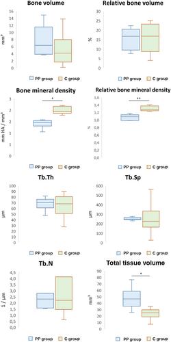
New mature lamellar bone was formed over the pristine bone in all groups. More intensive signs of bone modelling and remodelling were observed in the PP group compared to the C group. Mean new bone, bone marrow, connective tissue and total volumetric densities were greater in the PP group (p < 0.001, p = 0.001, p = 0.003 and p < 0.001, respectively). No differences were observed in the relative area parameters. Total tissue volume and bone volume were higher in the PP group (p = 0.031 and p = 0.076, respectively), while the bone mineral densities were higher in the C group (p = 0.041 and p = 0.003, respectively). Trabecular number, trabecular thickness and trabecular separation values were similar between the two groups.
Conclusions
Regeneration of vertical alveolar bone ridge defects may be enhanced by activation of the periosteum, without the application of bone grafting materials.
期刊介绍:
Journal of Clinical Periodontology was founded by the British, Dutch, French, German, Scandinavian, and Swiss Societies of Periodontology.
The aim of the Journal of Clinical Periodontology is to provide the platform for exchange of scientific and clinical progress in the field of Periodontology and allied disciplines, and to do so at the highest possible level. The Journal also aims to facilitate the application of new scientific knowledge to the daily practice of the concerned disciplines and addresses both practicing clinicians and academics. The Journal is the official publication of the European Federation of Periodontology but wishes to retain its international scope.
The Journal publishes original contributions of high scientific merit in the fields of periodontology and implant dentistry. Its scope encompasses the physiology and pathology of the periodontium, the tissue integration of dental implants, the biology and the modulation of periodontal and alveolar bone healing and regeneration, diagnosis, epidemiology, prevention and therapy of periodontal disease, the clinical aspects of tooth replacement with dental implants, and the comprehensive rehabilitation of the periodontal patient. Review articles by experts on new developments in basic and applied periodontal science and associated dental disciplines, advances in periodontal or implant techniques and procedures, and case reports which illustrate important new information are also welcome.
 求助内容:
求助内容: 应助结果提醒方式:


