Monahan Kevin, Hogan William, Matthew Chilton, Maher Michael, Hughes Alice, Altman Gregory, Altman Daniel, Hammarstedt Jon Erik
{"title":"骨盆经皮固定术的术中计算机断层扫描:回顾性病例系列。","authors":"Monahan Kevin, Hogan William, Matthew Chilton, Maher Michael, Hughes Alice, Altman Gregory, Altman Daniel, Hammarstedt Jon Erik","doi":"10.1007/s00264-024-06265-7","DOIUrl":null,"url":null,"abstract":"<p><strong>Purpose: </strong>Fractures and dislocations of the pelvic ring are complex injuries that when treating require meticulous attention to detail and often specialized technical skill. These injuries can be the result of high-energy trauma, particularly in younger patients, or low energy trauma more often found in the elderly. Regardless of mechanism, these injuries lie on a spectrum of severity and can be treated conservatively or surgically. Percutaneous fixation under fluoroscopic guidance is the preferred standard technique when treating these fractures. This technique can be challenging for a variety of reasons including patient characteristics, intra-operative image quality, fracture morphology, among others.</p><p><strong>Methods: </strong>This retrospective study evaluated the use of intra-operative computed tomography (CT) using an O-arm imaging system for critical evaluation of fluoroscopic-guided screw placement in twenty-three patients. We retrospectively reviewed all cases of patients who were treated by three fellowship-trained orthopaedic traumatologists during a one-year span. Patients undergoing percutaneous pelvis fixation using both standard fluoroscopy and intraoperative CT with the Medtronic O-arm® (Minneapolis, MN) imaging system. Additionally, procedures performed included open reduction internal fixation (ORIF) of the pelvic ring, acetabulum, and associated extremity fractures.</p><p><strong>Results: </strong>Twenty-three patients were included in this study. On average, the use of intraoperative CT added 24.4 min in operative time. Five patients (21.7%) required implant adjustment after O-arm spin. Fourteen patients underwent additional post-operative CT. No secondary revision surgeries were attempted after any post-operative CT.</p><p><strong>Conclusions: </strong>Our study suggests that intra-operative CT scan, compared to post-operative CT scan, can be utilized to prevent take-back surgery for misplaced implants and allow for adjustment in real-time.</p>","PeriodicalId":14450,"journal":{"name":"International Orthopaedics","volume":" ","pages":"2743-2748"},"PeriodicalIF":2.0000,"publicationDate":"2024-10-01","publicationTypes":"Journal Article","fieldsOfStudy":null,"isOpenAccess":false,"openAccessPdf":"https://www.ncbi.nlm.nih.gov/pmc/articles/PMC11422416/pdf/","citationCount":"0","resultStr":"{\"title\":\"Intraoperative computerised tomography scan for percutaneous fixation of the pelvis: a retrospective case series.\",\"authors\":\"Monahan Kevin, Hogan William, Matthew Chilton, Maher Michael, Hughes Alice, Altman Gregory, Altman Daniel, Hammarstedt Jon Erik\",\"doi\":\"10.1007/s00264-024-06265-7\",\"DOIUrl\":null,\"url\":null,\"abstract\":\"<p><strong>Purpose: </strong>Fractures and dislocations of the pelvic ring are complex injuries that when treating require meticulous attention to detail and often specialized technical skill. These injuries can be the result of high-energy trauma, particularly in younger patients, or low energy trauma more often found in the elderly. Regardless of mechanism, these injuries lie on a spectrum of severity and can be treated conservatively or surgically. Percutaneous fixation under fluoroscopic guidance is the preferred standard technique when treating these fractures. This technique can be challenging for a variety of reasons including patient characteristics, intra-operative image quality, fracture morphology, among others.</p><p><strong>Methods: </strong>This retrospective study evaluated the use of intra-operative computed tomography (CT) using an O-arm imaging system for critical evaluation of fluoroscopic-guided screw placement in twenty-three patients. We retrospectively reviewed all cases of patients who were treated by three fellowship-trained orthopaedic traumatologists during a one-year span. Patients undergoing percutaneous pelvis fixation using both standard fluoroscopy and intraoperative CT with the Medtronic O-arm® (Minneapolis, MN) imaging system. Additionally, procedures performed included open reduction internal fixation (ORIF) of the pelvic ring, acetabulum, and associated extremity fractures.</p><p><strong>Results: </strong>Twenty-three patients were included in this study. On average, the use of intraoperative CT added 24.4 min in operative time. Five patients (21.7%) required implant adjustment after O-arm spin. Fourteen patients underwent additional post-operative CT. No secondary revision surgeries were attempted after any post-operative CT.</p><p><strong>Conclusions: </strong>Our study suggests that intra-operative CT scan, compared to post-operative CT scan, can be utilized to prevent take-back surgery for misplaced implants and allow for adjustment in real-time.</p>\",\"PeriodicalId\":14450,\"journal\":{\"name\":\"International Orthopaedics\",\"volume\":\" \",\"pages\":\"2743-2748\"},\"PeriodicalIF\":2.0000,\"publicationDate\":\"2024-10-01\",\"publicationTypes\":\"Journal Article\",\"fieldsOfStudy\":null,\"isOpenAccess\":false,\"openAccessPdf\":\"https://www.ncbi.nlm.nih.gov/pmc/articles/PMC11422416/pdf/\",\"citationCount\":\"0\",\"resultStr\":null,\"platform\":\"Semanticscholar\",\"paperid\":null,\"PeriodicalName\":\"International Orthopaedics\",\"FirstCategoryId\":\"3\",\"ListUrlMain\":\"https://doi.org/10.1007/s00264-024-06265-7\",\"RegionNum\":3,\"RegionCategory\":\"医学\",\"ArticlePicture\":[],\"TitleCN\":null,\"AbstractTextCN\":null,\"PMCID\":null,\"EPubDate\":\"2024/8/15 0:00:00\",\"PubModel\":\"Epub\",\"JCR\":\"Q2\",\"JCRName\":\"ORTHOPEDICS\",\"Score\":null,\"Total\":0}","platform":"Semanticscholar","paperid":null,"PeriodicalName":"International Orthopaedics","FirstCategoryId":"3","ListUrlMain":"https://doi.org/10.1007/s00264-024-06265-7","RegionNum":3,"RegionCategory":"医学","ArticlePicture":[],"TitleCN":null,"AbstractTextCN":null,"PMCID":null,"EPubDate":"2024/8/15 0:00:00","PubModel":"Epub","JCR":"Q2","JCRName":"ORTHOPEDICS","Score":null,"Total":0}
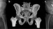
Intraoperative computerised tomography scan for percutaneous fixation of the pelvis: a retrospective case series.
Purpose: Fractures and dislocations of the pelvic ring are complex injuries that when treating require meticulous attention to detail and often specialized technical skill. These injuries can be the result of high-energy trauma, particularly in younger patients, or low energy trauma more often found in the elderly. Regardless of mechanism, these injuries lie on a spectrum of severity and can be treated conservatively or surgically. Percutaneous fixation under fluoroscopic guidance is the preferred standard technique when treating these fractures. This technique can be challenging for a variety of reasons including patient characteristics, intra-operative image quality, fracture morphology, among others.
Methods: This retrospective study evaluated the use of intra-operative computed tomography (CT) using an O-arm imaging system for critical evaluation of fluoroscopic-guided screw placement in twenty-three patients. We retrospectively reviewed all cases of patients who were treated by three fellowship-trained orthopaedic traumatologists during a one-year span. Patients undergoing percutaneous pelvis fixation using both standard fluoroscopy and intraoperative CT with the Medtronic O-arm® (Minneapolis, MN) imaging system. Additionally, procedures performed included open reduction internal fixation (ORIF) of the pelvic ring, acetabulum, and associated extremity fractures.
Results: Twenty-three patients were included in this study. On average, the use of intraoperative CT added 24.4 min in operative time. Five patients (21.7%) required implant adjustment after O-arm spin. Fourteen patients underwent additional post-operative CT. No secondary revision surgeries were attempted after any post-operative CT.
Conclusions: Our study suggests that intra-operative CT scan, compared to post-operative CT scan, can be utilized to prevent take-back surgery for misplaced implants and allow for adjustment in real-time.
期刊介绍:
International Orthopaedics, the Official Journal of the Société Internationale de Chirurgie Orthopédique et de Traumatologie (SICOT) , publishes original papers from all over the world. The articles deal with clinical orthopaedic surgery or basic research directly connected with orthopaedic surgery. International Orthopaedics will also link all the members of SICOT by means of an insert that will be concerned with SICOT matters.
Finally, it is expected that news and information regarding all aspects of orthopaedic surgery, including meetings, panels, instructional courses, etc. will be brought to the attention of the readers.
Manuscripts submitted for publication must contain a statement to the effect that all human studies have been approved by the appropriate ethics committee and have therefore been performed in accordance with the ethical standards laid down in the 1964 Declaration of Helsinki. It should also be stated clearly in the text that all persons gave their informed consent prior to their inclusion in the study. Details that might disclose the identity of the subjects under study should be omitted.
Reports of animal experiments must state that the "Principles of laboratory animal care" (NIH publication No. 85-23, revised 1985) were followed, as well as specific national laws (e.g. the current version of the German Law on the Protection of Animals) where applicable.
The editors reserve the right to reject manuscripts that do not comply with the above-mentioned requirements. The author will be held responsible for false statements or for failure to fulfil the above-mentioned requirements.
 求助内容:
求助内容: 应助结果提醒方式:


