Mi Zhou, Mengyuan Chen, Mingfang Luo, Meining Chen, Hongyun Huang
{"title":"基于扩散加权成像、体内非相干运动和扩散峰度成像的直肠癌病理预后因素。","authors":"Mi Zhou, Mengyuan Chen, Mingfang Luo, Meining Chen, Hongyun Huang","doi":"10.1007/s00330-024-11025-7","DOIUrl":null,"url":null,"abstract":"<p><strong>Objectives: </strong>To explore diffusion-weighted imaging (DWI), intravoxel incoherent motion (IVIM), and diffusion kurtosis imaging (DKI) for assessing pathological prognostic factors in patients with rectal cancer.</p><p><strong>Materials and methods: </strong>A total of 162 patients (105 males; mean age of 61.8 ± 13.1 years old) scheduled to undergo radical surgery were enrolled in this prospective study. The pathological prognostic factors included histological differentiation, lymph node metastasis (LNM), and extramural vascular invasion (EMVI). The DWI, IVIM, and DKI parameters were obtained and correlated with prognostic factors using univariable and multivariable logistic regression. Their assessment value was evaluated using receiver operating characteristic (ROC) curve analysis.</p><p><strong>Results: </strong>Multivariable logistic regression analyses showed that higher mean kurtosis (MK) (odds ratio (OR) = 194.931, p < 0.001) and lower apparent diffusion coefficient (ADC) (OR = 0.077, p = 0.025) were independently associated with poorer differentiation tumors. Higher perfusion fraction (f) (OR = 575.707, p = 0.023) and higher MK (OR = 173.559, p < 0.001) were independently associated with LNMs. Higher f (OR = 1036.116, p = 0.024), higher MK (OR = 253.629, p < 0.001), lower mean diffusivity (MD) (OR = 0.125, p = 0.038), and lower ADC (OR = 0.094, p = 0.022) were independently associated with EMVI. The area under the ROC curve (AUC) of MK for histological differentiation was significantly higher than ADC (0.771 vs. 0.638, p = 0.035). The AUC of MK for LNM positivity was higher than f (0.770 vs. 0.656, p = 0.048). The AUC of MK combined with MD (0.790) was the highest among f (0.663), MK (0.779), MD (0.617), and ADC (0.610) in assessing EMVI.</p><p><strong>Conclusion: </strong>The DKI parameters may be used as imaging biomarkers to assess pathological prognostic factors of rectal cancer before surgery.</p><p><strong>Clinical relevance statement: </strong>Diffusion kurtosis imaging (DKI) parameters, particularly mean kurtosis (MK), are promising biomarkers for assessing histological differentiation, lymph node metastasis, and extramural vascular invasion of rectal cancer. These findings suggest DKI's potential in the preoperative assessment of rectal cancer.</p><p><strong>Key points: </strong>Mean kurtosis outperformed the apparent diffusion coefficient in assessing histological differentiation in resectable rectal cancer. Perfusion fraction and mean kurtosis are independent indicators for assessing lymph node metastasis in rectal cancer. Mean kurtosis and mean diffusivity demonstrated superior accuracy in assessing extramural vascular invasion.</p>","PeriodicalId":12076,"journal":{"name":"European Radiology","volume":" ","pages":"979-988"},"PeriodicalIF":4.7000,"publicationDate":"2025-02-01","publicationTypes":"Journal Article","fieldsOfStudy":null,"isOpenAccess":false,"openAccessPdf":"","citationCount":"0","resultStr":"{\"title\":\"Pathological prognostic factors of rectal cancer based on diffusion-weighted imaging, intravoxel incoherent motion, and diffusion kurtosis imaging.\",\"authors\":\"Mi Zhou, Mengyuan Chen, Mingfang Luo, Meining Chen, Hongyun Huang\",\"doi\":\"10.1007/s00330-024-11025-7\",\"DOIUrl\":null,\"url\":null,\"abstract\":\"<p><strong>Objectives: </strong>To explore diffusion-weighted imaging (DWI), intravoxel incoherent motion (IVIM), and diffusion kurtosis imaging (DKI) for assessing pathological prognostic factors in patients with rectal cancer.</p><p><strong>Materials and methods: </strong>A total of 162 patients (105 males; mean age of 61.8 ± 13.1 years old) scheduled to undergo radical surgery were enrolled in this prospective study. The pathological prognostic factors included histological differentiation, lymph node metastasis (LNM), and extramural vascular invasion (EMVI). The DWI, IVIM, and DKI parameters were obtained and correlated with prognostic factors using univariable and multivariable logistic regression. Their assessment value was evaluated using receiver operating characteristic (ROC) curve analysis.</p><p><strong>Results: </strong>Multivariable logistic regression analyses showed that higher mean kurtosis (MK) (odds ratio (OR) = 194.931, p < 0.001) and lower apparent diffusion coefficient (ADC) (OR = 0.077, p = 0.025) were independently associated with poorer differentiation tumors. Higher perfusion fraction (f) (OR = 575.707, p = 0.023) and higher MK (OR = 173.559, p < 0.001) were independently associated with LNMs. Higher f (OR = 1036.116, p = 0.024), higher MK (OR = 253.629, p < 0.001), lower mean diffusivity (MD) (OR = 0.125, p = 0.038), and lower ADC (OR = 0.094, p = 0.022) were independently associated with EMVI. The area under the ROC curve (AUC) of MK for histological differentiation was significantly higher than ADC (0.771 vs. 0.638, p = 0.035). The AUC of MK for LNM positivity was higher than f (0.770 vs. 0.656, p = 0.048). The AUC of MK combined with MD (0.790) was the highest among f (0.663), MK (0.779), MD (0.617), and ADC (0.610) in assessing EMVI.</p><p><strong>Conclusion: </strong>The DKI parameters may be used as imaging biomarkers to assess pathological prognostic factors of rectal cancer before surgery.</p><p><strong>Clinical relevance statement: </strong>Diffusion kurtosis imaging (DKI) parameters, particularly mean kurtosis (MK), are promising biomarkers for assessing histological differentiation, lymph node metastasis, and extramural vascular invasion of rectal cancer. These findings suggest DKI's potential in the preoperative assessment of rectal cancer.</p><p><strong>Key points: </strong>Mean kurtosis outperformed the apparent diffusion coefficient in assessing histological differentiation in resectable rectal cancer. Perfusion fraction and mean kurtosis are independent indicators for assessing lymph node metastasis in rectal cancer. Mean kurtosis and mean diffusivity demonstrated superior accuracy in assessing extramural vascular invasion.</p>\",\"PeriodicalId\":12076,\"journal\":{\"name\":\"European Radiology\",\"volume\":\" \",\"pages\":\"979-988\"},\"PeriodicalIF\":4.7000,\"publicationDate\":\"2025-02-01\",\"publicationTypes\":\"Journal Article\",\"fieldsOfStudy\":null,\"isOpenAccess\":false,\"openAccessPdf\":\"\",\"citationCount\":\"0\",\"resultStr\":null,\"platform\":\"Semanticscholar\",\"paperid\":null,\"PeriodicalName\":\"European Radiology\",\"FirstCategoryId\":\"3\",\"ListUrlMain\":\"https://doi.org/10.1007/s00330-024-11025-7\",\"RegionNum\":2,\"RegionCategory\":\"医学\",\"ArticlePicture\":[],\"TitleCN\":null,\"AbstractTextCN\":null,\"PMCID\":null,\"EPubDate\":\"2024/8/14 0:00:00\",\"PubModel\":\"Epub\",\"JCR\":\"Q1\",\"JCRName\":\"RADIOLOGY, NUCLEAR MEDICINE & MEDICAL IMAGING\",\"Score\":null,\"Total\":0}","platform":"Semanticscholar","paperid":null,"PeriodicalName":"European Radiology","FirstCategoryId":"3","ListUrlMain":"https://doi.org/10.1007/s00330-024-11025-7","RegionNum":2,"RegionCategory":"医学","ArticlePicture":[],"TitleCN":null,"AbstractTextCN":null,"PMCID":null,"EPubDate":"2024/8/14 0:00:00","PubModel":"Epub","JCR":"Q1","JCRName":"RADIOLOGY, NUCLEAR MEDICINE & MEDICAL IMAGING","Score":null,"Total":0}
Pathological prognostic factors of rectal cancer based on diffusion-weighted imaging, intravoxel incoherent motion, and diffusion kurtosis imaging.
Objectives: To explore diffusion-weighted imaging (DWI), intravoxel incoherent motion (IVIM), and diffusion kurtosis imaging (DKI) for assessing pathological prognostic factors in patients with rectal cancer.
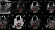
Materials and methods: A total of 162 patients (105 males; mean age of 61.8 ± 13.1 years old) scheduled to undergo radical surgery were enrolled in this prospective study. The pathological prognostic factors included histological differentiation, lymph node metastasis (LNM), and extramural vascular invasion (EMVI). The DWI, IVIM, and DKI parameters were obtained and correlated with prognostic factors using univariable and multivariable logistic regression. Their assessment value was evaluated using receiver operating characteristic (ROC) curve analysis.
Results: Multivariable logistic regression analyses showed that higher mean kurtosis (MK) (odds ratio (OR) = 194.931, p < 0.001) and lower apparent diffusion coefficient (ADC) (OR = 0.077, p = 0.025) were independently associated with poorer differentiation tumors. Higher perfusion fraction (f) (OR = 575.707, p = 0.023) and higher MK (OR = 173.559, p < 0.001) were independently associated with LNMs. Higher f (OR = 1036.116, p = 0.024), higher MK (OR = 253.629, p < 0.001), lower mean diffusivity (MD) (OR = 0.125, p = 0.038), and lower ADC (OR = 0.094, p = 0.022) were independently associated with EMVI. The area under the ROC curve (AUC) of MK for histological differentiation was significantly higher than ADC (0.771 vs. 0.638, p = 0.035). The AUC of MK for LNM positivity was higher than f (0.770 vs. 0.656, p = 0.048). The AUC of MK combined with MD (0.790) was the highest among f (0.663), MK (0.779), MD (0.617), and ADC (0.610) in assessing EMVI.
Conclusion: The DKI parameters may be used as imaging biomarkers to assess pathological prognostic factors of rectal cancer before surgery.
Clinical relevance statement: Diffusion kurtosis imaging (DKI) parameters, particularly mean kurtosis (MK), are promising biomarkers for assessing histological differentiation, lymph node metastasis, and extramural vascular invasion of rectal cancer. These findings suggest DKI's potential in the preoperative assessment of rectal cancer.
Key points: Mean kurtosis outperformed the apparent diffusion coefficient in assessing histological differentiation in resectable rectal cancer. Perfusion fraction and mean kurtosis are independent indicators for assessing lymph node metastasis in rectal cancer. Mean kurtosis and mean diffusivity demonstrated superior accuracy in assessing extramural vascular invasion.
期刊介绍:
European Radiology (ER) continuously updates scientific knowledge in radiology by publication of strong original articles and state-of-the-art reviews written by leading radiologists. A well balanced combination of review articles, original papers, short communications from European radiological congresses and information on society matters makes ER an indispensable source for current information in this field.
This is the Journal of the European Society of Radiology, and the official journal of a number of societies.
From 2004-2008 supplements to European Radiology were published under its companion, European Radiology Supplements, ISSN 1613-3749.

 求助内容:
求助内容: 应助结果提醒方式:


