Marcin Wrzosek, Aleksandra Banasik, Adriana Czerwik, Agnieszka Olszewska, Marta Płonek, Veronika Stein
{"title":"在癫痫犬中使用镇静-唤醒脑电图。","authors":"Marcin Wrzosek, Aleksandra Banasik, Adriana Czerwik, Agnieszka Olszewska, Marta Płonek, Veronika Stein","doi":"10.1111/jvim.17153","DOIUrl":null,"url":null,"abstract":"<div>\n \n \n <section>\n \n <h3> Background</h3>\n \n <p>Electroencephalography (EEG) recording protocols have been standardized for humans. Although the utilization of techniques in veterinary medicine is increasing, a standard protocol has not yet been established.</p>\n </section>\n \n <section>\n \n <h3> Hypothesis</h3>\n \n <p>Assessment of a sedation-awakening EEG protocol in dogs.</p>\n </section>\n \n <section>\n \n <h3> Animals</h3>\n \n <p>Electroencephalography examination was performed in a research colony of 6 nonepileptic dogs (control [C]) and 12 dogs with epilepsy admitted to the clinic because of the epileptic seizures.</p>\n </section>\n \n <section>\n \n <h3> Methods</h3>\n \n <p>It was a prospective study with retrospective control. Dogs with epilepsy were divided into 2 equal groups, wherein EEG acquisition was performed using a “sedation” protocol (IE-S, n = 6) and a “sedation-awakening” protocol (IE-SA, n = 6). All animals were sedated using medetomidine. In IE-SA group, sedation was reversed 5 minutes after commencing the EEG recording by injecting atipamezole IM. Type of background activity (BGA) and presence of EEG-defined epileptiform discharges (EDs) were evaluated blindly. Statistical significance was set at P > 0.05.</p>\n </section>\n \n <section>\n \n <h3> Results</h3>\n \n <p>Epileptiform discharges were found in 1 of 6 of the dogs in group C, 4 of 6 of the dogs in IE-S group, and 5 of 6 of the dogs in IE-SA group. A significantly greater number of EDs (spikes, <i>P</i> = .0109; polyspikes, <i>P</i> = .0109; sharp waves, <i>P</i> = .01) were detected in Phase 2 in animals subjected to the “sedation-awakening” protocol, whereas there was no statistically significant greater number of discharges in sedated animals.</p>\n </section>\n \n <section>\n \n <h3> Conclusions and Clinical Importance</h3>\n \n <p>A “sedation-awakening” EEG protocol could be of value for ambulatory use if repeated EEG recordings and monitoring of epilepsy in dogs is needed.</p>\n </section>\n </div>","PeriodicalId":49958,"journal":{"name":"Journal of Veterinary Internal Medicine","volume":null,"pages":null},"PeriodicalIF":2.1000,"publicationDate":"2024-08-12","publicationTypes":"Journal Article","fieldsOfStudy":null,"isOpenAccess":false,"openAccessPdf":"https://onlinelibrary.wiley.com/doi/epdf/10.1111/jvim.17153","citationCount":"0","resultStr":"{\"title\":\"Use of sedation-awakening electroencephalography in dogs with epilepsy\",\"authors\":\"Marcin Wrzosek, Aleksandra Banasik, Adriana Czerwik, Agnieszka Olszewska, Marta Płonek, Veronika Stein\",\"doi\":\"10.1111/jvim.17153\",\"DOIUrl\":null,\"url\":null,\"abstract\":\"<div>\\n \\n \\n <section>\\n \\n <h3> Background</h3>\\n \\n <p>Electroencephalography (EEG) recording protocols have been standardized for humans. Although the utilization of techniques in veterinary medicine is increasing, a standard protocol has not yet been established.</p>\\n </section>\\n \\n <section>\\n \\n <h3> Hypothesis</h3>\\n \\n <p>Assessment of a sedation-awakening EEG protocol in dogs.</p>\\n </section>\\n \\n <section>\\n \\n <h3> Animals</h3>\\n \\n <p>Electroencephalography examination was performed in a research colony of 6 nonepileptic dogs (control [C]) and 12 dogs with epilepsy admitted to the clinic because of the epileptic seizures.</p>\\n </section>\\n \\n <section>\\n \\n <h3> Methods</h3>\\n \\n <p>It was a prospective study with retrospective control. Dogs with epilepsy were divided into 2 equal groups, wherein EEG acquisition was performed using a “sedation” protocol (IE-S, n = 6) and a “sedation-awakening” protocol (IE-SA, n = 6). All animals were sedated using medetomidine. In IE-SA group, sedation was reversed 5 minutes after commencing the EEG recording by injecting atipamezole IM. Type of background activity (BGA) and presence of EEG-defined epileptiform discharges (EDs) were evaluated blindly. Statistical significance was set at P > 0.05.</p>\\n </section>\\n \\n <section>\\n \\n <h3> Results</h3>\\n \\n <p>Epileptiform discharges were found in 1 of 6 of the dogs in group C, 4 of 6 of the dogs in IE-S group, and 5 of 6 of the dogs in IE-SA group. A significantly greater number of EDs (spikes, <i>P</i> = .0109; polyspikes, <i>P</i> = .0109; sharp waves, <i>P</i> = .01) were detected in Phase 2 in animals subjected to the “sedation-awakening” protocol, whereas there was no statistically significant greater number of discharges in sedated animals.</p>\\n </section>\\n \\n <section>\\n \\n <h3> Conclusions and Clinical Importance</h3>\\n \\n <p>A “sedation-awakening” EEG protocol could be of value for ambulatory use if repeated EEG recordings and monitoring of epilepsy in dogs is needed.</p>\\n </section>\\n </div>\",\"PeriodicalId\":49958,\"journal\":{\"name\":\"Journal of Veterinary Internal Medicine\",\"volume\":null,\"pages\":null},\"PeriodicalIF\":2.1000,\"publicationDate\":\"2024-08-12\",\"publicationTypes\":\"Journal Article\",\"fieldsOfStudy\":null,\"isOpenAccess\":false,\"openAccessPdf\":\"https://onlinelibrary.wiley.com/doi/epdf/10.1111/jvim.17153\",\"citationCount\":\"0\",\"resultStr\":null,\"platform\":\"Semanticscholar\",\"paperid\":null,\"PeriodicalName\":\"Journal of Veterinary Internal Medicine\",\"FirstCategoryId\":\"97\",\"ListUrlMain\":\"https://onlinelibrary.wiley.com/doi/10.1111/jvim.17153\",\"RegionNum\":2,\"RegionCategory\":\"农林科学\",\"ArticlePicture\":[],\"TitleCN\":null,\"AbstractTextCN\":null,\"PMCID\":null,\"EPubDate\":\"\",\"PubModel\":\"\",\"JCR\":\"Q1\",\"JCRName\":\"VETERINARY SCIENCES\",\"Score\":null,\"Total\":0}","platform":"Semanticscholar","paperid":null,"PeriodicalName":"Journal of Veterinary Internal Medicine","FirstCategoryId":"97","ListUrlMain":"https://onlinelibrary.wiley.com/doi/10.1111/jvim.17153","RegionNum":2,"RegionCategory":"农林科学","ArticlePicture":[],"TitleCN":null,"AbstractTextCN":null,"PMCID":null,"EPubDate":"","PubModel":"","JCR":"Q1","JCRName":"VETERINARY SCIENCES","Score":null,"Total":0}
Use of sedation-awakening electroencephalography in dogs with epilepsy
Background
Electroencephalography (EEG) recording protocols have been standardized for humans. Although the utilization of techniques in veterinary medicine is increasing, a standard protocol has not yet been established.
Hypothesis
Assessment of a sedation-awakening EEG protocol in dogs.
Animals
Electroencephalography examination was performed in a research colony of 6 nonepileptic dogs (control [C]) and 12 dogs with epilepsy admitted to the clinic because of the epileptic seizures.
Methods
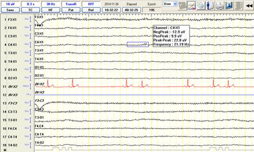
It was a prospective study with retrospective control. Dogs with epilepsy were divided into 2 equal groups, wherein EEG acquisition was performed using a “sedation” protocol (IE-S, n = 6) and a “sedation-awakening” protocol (IE-SA, n = 6). All animals were sedated using medetomidine. In IE-SA group, sedation was reversed 5 minutes after commencing the EEG recording by injecting atipamezole IM. Type of background activity (BGA) and presence of EEG-defined epileptiform discharges (EDs) were evaluated blindly. Statistical significance was set at P > 0.05.
Results
Epileptiform discharges were found in 1 of 6 of the dogs in group C, 4 of 6 of the dogs in IE-S group, and 5 of 6 of the dogs in IE-SA group. A significantly greater number of EDs (spikes, P = .0109; polyspikes, P = .0109; sharp waves, P = .01) were detected in Phase 2 in animals subjected to the “sedation-awakening” protocol, whereas there was no statistically significant greater number of discharges in sedated animals.
Conclusions and Clinical Importance
A “sedation-awakening” EEG protocol could be of value for ambulatory use if repeated EEG recordings and monitoring of epilepsy in dogs is needed.
期刊介绍:
The mission of the Journal of Veterinary Internal Medicine is to advance veterinary medical knowledge and improve the lives of animals by publication of authoritative scientific articles of animal diseases.
 求助内容:
求助内容: 应助结果提醒方式:


