用 82Rb PET 对心肌血流进行参数成像:准确性和图像质量分析
IF 3
4区 医学
Q2 CARDIAC & CARDIOVASCULAR SYSTEMS
引用次数: 0
摘要
背景:我们旨在开发一种生成三维心肌血流(MBF)图像的框架,对照临床验证的左心室二维极性 MBF 图计算其准确性,并评估其与相对心肌灌注成像(MPI)相比在图像质量方面的改进:回顾性研究了 N=40 名患者,他们的缺损严重程度和摄取动态各不相同。FlowQuantTM软件用于生成参考MPI和极性MBF图,并适用于体素MBF图。我们评估了参数值与极值之间在静息和应激时 MBF 以及储备(应激/静息 MBF)方面的一致性。我们还评估了从相对 MPI 到 MBF 的图像质量改善情况,评估指标包括信噪比、对比度-信噪比、组织-血液比和缺损严重程度:所有血流参数的三维参数图和二维极坐标图之间的一致性非常好(ICC>0.96),尽管偏差很小(ICC>0.96)。本文章由计算机程序翻译,如有差异,请以英文原文为准。
Parametric imaging of myocardial blood flow with 82Rb PET: An accuracy and image quality analysis
Background
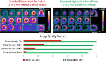
We aimed to develop a framework for generating three-dimensional (3D) myocardial blood flow (MBF) images, computing their accuracy against clinically validated two-dimensional (2D) polar MBF maps of the left ventricle, and evaluating their improvements in image quality over relative myocardial perfusion imaging (MPI).
Methods
N = 40 patients with a wide range of defect severities and uptake dynamics were retrospectively studied. The FlowQuant™ software was used to generate reference MPI and polar MBF maps and was adapted for voxel-wise MBF mapping. We evaluated agreement between parametric vs polar values for MBF at rest and stress and for reserve (stress/rest MBF). We also assessed improvements in image quality, assessed by signal-to-noise ratio, contrast-to-noise ratio, tissue-to-blood ratio, and defect severity, from relative MPI to MBF.
Results
There was excellent agreement between 3D parametric and 2D polar maps for all flow parameters (interclass correlation coefficient >0.96), albeit with minimal bias (<8%) for rest and stress MBF at the patient level. Image quality substantially improved from MPI to MBF in every patient for all image-quality metrics (P < 0.0001)
Conclusions
We developed a robust methodology for producing highly accurate 3D MBF images exhibiting considerably improved image quality compared to relative MPI commonly used in clinical practice.
求助全文
通过发布文献求助,成功后即可免费获取论文全文。
去求助
来源期刊
CiteScore
5.30
自引率
20.80%
发文量
249
审稿时长
4-8 weeks
期刊介绍:
Journal of Nuclear Cardiology is the only journal in the world devoted to this dynamic and growing subspecialty. Physicians and technologists value the Journal not only for its peer-reviewed articles, but also for its timely discussions about the current and future role of nuclear cardiology. Original articles address all aspects of nuclear cardiology, including interpretation, diagnosis, imaging equipment, and use of radiopharmaceuticals. As the official publication of the American Society of Nuclear Cardiology, the Journal also brings readers the latest information emerging from the Society''s task forces and publishes guidelines and position papers as they are adopted.
 求助内容:
求助内容: 应助结果提醒方式:


