Saumya Shivakumar, Kausalya K Sahu, Ranjitha Rao, Chaithra Gv, Cheryl Sarah Philipose, Sharada Rai
{"title":"ER、p53、CEA 和 Napsin A 在子宫内膜癌组织学亚型中的实用性及其与临床病理预后参数的相关性:一家转诊机构的经验。","authors":"Saumya Shivakumar, Kausalya K Sahu, Ranjitha Rao, Chaithra Gv, Cheryl Sarah Philipose, Sharada Rai","doi":"10.30699/IJP.2024.2008693.3154","DOIUrl":null,"url":null,"abstract":"<p><strong>Background & objective: </strong>Endometrial Carcinoma (EC) is the most common gynecological cancer with a global incidence of 23.2 per 1 lakh population. Histological subclassification of EC is extremely crucial for the diagnosis, proper management strategies, and prognosis. This study was conducted in a tertiary care institute to analyze the expression pattern of a minimum panel of 4 markers (ER, p53, CEA, Napsin A) with emphasis on their utility in the routine histological subtyping, aberrant expression, and correlation with various clinicopathological parameters.</p><p><strong>Methods: </strong>A time-bound cross-sectional observational and analytical study was conducted, which includes cases diagnosed in our laboratory from January 2016 to April 2021.</p><p><strong>Results: </strong>Sixty cases diagnosed as EC during the study period formed the sample cases. The ER was expressed in 85% (53/60) of cases in the current study. Among them, 94% (50/53) were endometrioid endometrial carcinomas (EECs). A negative correlation was found between ER intensity and age (r= -1.48). Of 60 EC cases, 10 (16%) cases expressed p53. The tumors positive for p53 with higher intensity were negative for ER and vice versa. The expression pattern of ER and p53 was statistically significant (<i>P</i>=-0.021). On IHC, 84.6% (11/13) of CEA-positive cases expressed both ER and CEA, suggesting mucinous differentiation. Napsin A was expressed in two cases of EEC, FIGO grade I, and one case of serous carcinoma.</p><p><strong>Conclusion: </strong>An inverse association was found between ER and p53 expression. The CEA is valuable in identifying EEC with mucinous differentiation.</p>","PeriodicalId":38900,"journal":{"name":"Iranian Journal of Pathology","volume":"19 2","pages":"236-243"},"PeriodicalIF":0.0000,"publicationDate":"2024-01-01","publicationTypes":"Journal Article","fieldsOfStudy":null,"isOpenAccess":false,"openAccessPdf":"https://www.ncbi.nlm.nih.gov/pmc/articles/PMC11304467/pdf/","citationCount":"0","resultStr":"{\"title\":\"Utility of ER, p53, CEA and Napsin A in Histological Subtyping of Endometrial Carcinoma and Their Correlation with Clinicopathological Prognostic Parameters: Experience from a Referral Institute.\",\"authors\":\"Saumya Shivakumar, Kausalya K Sahu, Ranjitha Rao, Chaithra Gv, Cheryl Sarah Philipose, Sharada Rai\",\"doi\":\"10.30699/IJP.2024.2008693.3154\",\"DOIUrl\":null,\"url\":null,\"abstract\":\"<p><strong>Background & objective: </strong>Endometrial Carcinoma (EC) is the most common gynecological cancer with a global incidence of 23.2 per 1 lakh population. Histological subclassification of EC is extremely crucial for the diagnosis, proper management strategies, and prognosis. This study was conducted in a tertiary care institute to analyze the expression pattern of a minimum panel of 4 markers (ER, p53, CEA, Napsin A) with emphasis on their utility in the routine histological subtyping, aberrant expression, and correlation with various clinicopathological parameters.</p><p><strong>Methods: </strong>A time-bound cross-sectional observational and analytical study was conducted, which includes cases diagnosed in our laboratory from January 2016 to April 2021.</p><p><strong>Results: </strong>Sixty cases diagnosed as EC during the study period formed the sample cases. The ER was expressed in 85% (53/60) of cases in the current study. Among them, 94% (50/53) were endometrioid endometrial carcinomas (EECs). A negative correlation was found between ER intensity and age (r= -1.48). Of 60 EC cases, 10 (16%) cases expressed p53. The tumors positive for p53 with higher intensity were negative for ER and vice versa. The expression pattern of ER and p53 was statistically significant (<i>P</i>=-0.021). On IHC, 84.6% (11/13) of CEA-positive cases expressed both ER and CEA, suggesting mucinous differentiation. Napsin A was expressed in two cases of EEC, FIGO grade I, and one case of serous carcinoma.</p><p><strong>Conclusion: </strong>An inverse association was found between ER and p53 expression. The CEA is valuable in identifying EEC with mucinous differentiation.</p>\",\"PeriodicalId\":38900,\"journal\":{\"name\":\"Iranian Journal of Pathology\",\"volume\":\"19 2\",\"pages\":\"236-243\"},\"PeriodicalIF\":0.0000,\"publicationDate\":\"2024-01-01\",\"publicationTypes\":\"Journal Article\",\"fieldsOfStudy\":null,\"isOpenAccess\":false,\"openAccessPdf\":\"https://www.ncbi.nlm.nih.gov/pmc/articles/PMC11304467/pdf/\",\"citationCount\":\"0\",\"resultStr\":null,\"platform\":\"Semanticscholar\",\"paperid\":null,\"PeriodicalName\":\"Iranian Journal of Pathology\",\"FirstCategoryId\":\"1085\",\"ListUrlMain\":\"https://doi.org/10.30699/IJP.2024.2008693.3154\",\"RegionNum\":0,\"RegionCategory\":null,\"ArticlePicture\":[],\"TitleCN\":null,\"AbstractTextCN\":null,\"PMCID\":null,\"EPubDate\":\"2024/1/29 0:00:00\",\"PubModel\":\"Epub\",\"JCR\":\"Q3\",\"JCRName\":\"Medicine\",\"Score\":null,\"Total\":0}","platform":"Semanticscholar","paperid":null,"PeriodicalName":"Iranian Journal of Pathology","FirstCategoryId":"1085","ListUrlMain":"https://doi.org/10.30699/IJP.2024.2008693.3154","RegionNum":0,"RegionCategory":null,"ArticlePicture":[],"TitleCN":null,"AbstractTextCN":null,"PMCID":null,"EPubDate":"2024/1/29 0:00:00","PubModel":"Epub","JCR":"Q3","JCRName":"Medicine","Score":null,"Total":0}
Utility of ER, p53, CEA and Napsin A in Histological Subtyping of Endometrial Carcinoma and Their Correlation with Clinicopathological Prognostic Parameters: Experience from a Referral Institute.
Background & objective: Endometrial Carcinoma (EC) is the most common gynecological cancer with a global incidence of 23.2 per 1 lakh population. Histological subclassification of EC is extremely crucial for the diagnosis, proper management strategies, and prognosis. This study was conducted in a tertiary care institute to analyze the expression pattern of a minimum panel of 4 markers (ER, p53, CEA, Napsin A) with emphasis on their utility in the routine histological subtyping, aberrant expression, and correlation with various clinicopathological parameters.
Methods: A time-bound cross-sectional observational and analytical study was conducted, which includes cases diagnosed in our laboratory from January 2016 to April 2021.
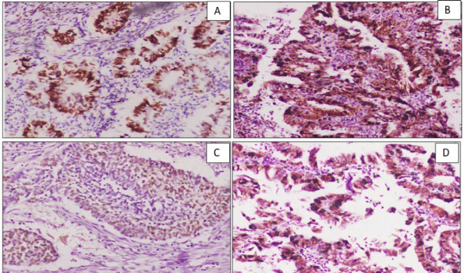
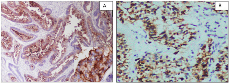
Results: Sixty cases diagnosed as EC during the study period formed the sample cases. The ER was expressed in 85% (53/60) of cases in the current study. Among them, 94% (50/53) were endometrioid endometrial carcinomas (EECs). A negative correlation was found between ER intensity and age (r= -1.48). Of 60 EC cases, 10 (16%) cases expressed p53. The tumors positive for p53 with higher intensity were negative for ER and vice versa. The expression pattern of ER and p53 was statistically significant (P=-0.021). On IHC, 84.6% (11/13) of CEA-positive cases expressed both ER and CEA, suggesting mucinous differentiation. Napsin A was expressed in two cases of EEC, FIGO grade I, and one case of serous carcinoma.
Conclusion: An inverse association was found between ER and p53 expression. The CEA is valuable in identifying EEC with mucinous differentiation.

 求助内容:
求助内容: 应助结果提醒方式:


