Ramsha Ahmed, Aamna Al Shehhi, Naoufel Werghi, Mohamed L. Seghier
{"title":"利用变压器增强核磁共振成像分析对中风病灶进行分割。","authors":"Ramsha Ahmed, Aamna Al Shehhi, Naoufel Werghi, Mohamed L. Seghier","doi":"10.1002/hbm.26803","DOIUrl":null,"url":null,"abstract":"<p>Accurate segmentation of chronic stroke lesions from mono-spectral magnetic resonance imaging scans (e.g., T1-weighted images) is a difficult task due to the arbitrary shape, complex texture, variable size and intensities, and varied locations of the lesions. Due to this inherent spatial heterogeneity, existing machine learning methods have shown moderate performance for chronic lesion delineation. In this study, we introduced: (1) a method that integrates transformers' deformable feature attention mechanism with convolutional deep learning architecture to improve the accuracy and generalizability of stroke lesion segmentation, and (2) an ecological data augmentation technique based on inserting real lesions into intact brain regions. Our combination of these two approaches resulted in a significant increase in segmentation performance, with a Dice index of 0.82 (±0.39), outperforming the existing methods trained and tested on the same Anatomical Tracings of Lesions After Stroke (ATLAS) 2022 dataset. Our method performed relatively well even for cases with small stroke lesions. We validated the robustness of our method through an ablation study and by testing it on new unseen brain scans from the Ischemic Stroke Lesion Segmentation (ISLES) 2015 dataset. Overall, our proposed approach of transformers with ecological data augmentation offers a robust way to delineate chronic stroke lesions with clinically relevant accuracy. Our method can be extended to other challenging tasks that require automated detection and segmentation of diverse brain abnormalities from clinical scans.</p>","PeriodicalId":13019,"journal":{"name":"Human Brain Mapping","volume":"45 11","pages":""},"PeriodicalIF":3.3000,"publicationDate":"2024-08-09","publicationTypes":"Journal Article","fieldsOfStudy":null,"isOpenAccess":false,"openAccessPdf":"https://www.ncbi.nlm.nih.gov/pmc/articles/PMC11310771/pdf/","citationCount":"0","resultStr":"{\"title\":\"Segmentation of stroke lesions using transformers-augmented MRI analysis\",\"authors\":\"Ramsha Ahmed, Aamna Al Shehhi, Naoufel Werghi, Mohamed L. Seghier\",\"doi\":\"10.1002/hbm.26803\",\"DOIUrl\":null,\"url\":null,\"abstract\":\"<p>Accurate segmentation of chronic stroke lesions from mono-spectral magnetic resonance imaging scans (e.g., T1-weighted images) is a difficult task due to the arbitrary shape, complex texture, variable size and intensities, and varied locations of the lesions. Due to this inherent spatial heterogeneity, existing machine learning methods have shown moderate performance for chronic lesion delineation. In this study, we introduced: (1) a method that integrates transformers' deformable feature attention mechanism with convolutional deep learning architecture to improve the accuracy and generalizability of stroke lesion segmentation, and (2) an ecological data augmentation technique based on inserting real lesions into intact brain regions. Our combination of these two approaches resulted in a significant increase in segmentation performance, with a Dice index of 0.82 (±0.39), outperforming the existing methods trained and tested on the same Anatomical Tracings of Lesions After Stroke (ATLAS) 2022 dataset. Our method performed relatively well even for cases with small stroke lesions. We validated the robustness of our method through an ablation study and by testing it on new unseen brain scans from the Ischemic Stroke Lesion Segmentation (ISLES) 2015 dataset. Overall, our proposed approach of transformers with ecological data augmentation offers a robust way to delineate chronic stroke lesions with clinically relevant accuracy. Our method can be extended to other challenging tasks that require automated detection and segmentation of diverse brain abnormalities from clinical scans.</p>\",\"PeriodicalId\":13019,\"journal\":{\"name\":\"Human Brain Mapping\",\"volume\":\"45 11\",\"pages\":\"\"},\"PeriodicalIF\":3.3000,\"publicationDate\":\"2024-08-09\",\"publicationTypes\":\"Journal Article\",\"fieldsOfStudy\":null,\"isOpenAccess\":false,\"openAccessPdf\":\"https://www.ncbi.nlm.nih.gov/pmc/articles/PMC11310771/pdf/\",\"citationCount\":\"0\",\"resultStr\":null,\"platform\":\"Semanticscholar\",\"paperid\":null,\"PeriodicalName\":\"Human Brain Mapping\",\"FirstCategoryId\":\"3\",\"ListUrlMain\":\"https://onlinelibrary.wiley.com/doi/10.1002/hbm.26803\",\"RegionNum\":2,\"RegionCategory\":\"医学\",\"ArticlePicture\":[],\"TitleCN\":null,\"AbstractTextCN\":null,\"PMCID\":null,\"EPubDate\":\"\",\"PubModel\":\"\",\"JCR\":\"Q1\",\"JCRName\":\"NEUROIMAGING\",\"Score\":null,\"Total\":0}","platform":"Semanticscholar","paperid":null,"PeriodicalName":"Human Brain Mapping","FirstCategoryId":"3","ListUrlMain":"https://onlinelibrary.wiley.com/doi/10.1002/hbm.26803","RegionNum":2,"RegionCategory":"医学","ArticlePicture":[],"TitleCN":null,"AbstractTextCN":null,"PMCID":null,"EPubDate":"","PubModel":"","JCR":"Q1","JCRName":"NEUROIMAGING","Score":null,"Total":0}
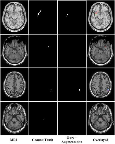
Segmentation of stroke lesions using transformers-augmented MRI analysis
Accurate segmentation of chronic stroke lesions from mono-spectral magnetic resonance imaging scans (e.g., T1-weighted images) is a difficult task due to the arbitrary shape, complex texture, variable size and intensities, and varied locations of the lesions. Due to this inherent spatial heterogeneity, existing machine learning methods have shown moderate performance for chronic lesion delineation. In this study, we introduced: (1) a method that integrates transformers' deformable feature attention mechanism with convolutional deep learning architecture to improve the accuracy and generalizability of stroke lesion segmentation, and (2) an ecological data augmentation technique based on inserting real lesions into intact brain regions. Our combination of these two approaches resulted in a significant increase in segmentation performance, with a Dice index of 0.82 (±0.39), outperforming the existing methods trained and tested on the same Anatomical Tracings of Lesions After Stroke (ATLAS) 2022 dataset. Our method performed relatively well even for cases with small stroke lesions. We validated the robustness of our method through an ablation study and by testing it on new unseen brain scans from the Ischemic Stroke Lesion Segmentation (ISLES) 2015 dataset. Overall, our proposed approach of transformers with ecological data augmentation offers a robust way to delineate chronic stroke lesions with clinically relevant accuracy. Our method can be extended to other challenging tasks that require automated detection and segmentation of diverse brain abnormalities from clinical scans.
期刊介绍:
Human Brain Mapping publishes peer-reviewed basic, clinical, technical, and theoretical research in the interdisciplinary and rapidly expanding field of human brain mapping. The journal features research derived from non-invasive brain imaging modalities used to explore the spatial and temporal organization of the neural systems supporting human behavior. Imaging modalities of interest include positron emission tomography, event-related potentials, electro-and magnetoencephalography, magnetic resonance imaging, and single-photon emission tomography. Brain mapping research in both normal and clinical populations is encouraged.
Article formats include Research Articles, Review Articles, Clinical Case Studies, and Technique, as well as Technological Developments, Theoretical Articles, and Synthetic Reviews. Technical advances, such as novel brain imaging methods, analyses for detecting or localizing neural activity, synergistic uses of multiple imaging modalities, and strategies for the design of behavioral paradigms and neural-systems modeling are of particular interest. The journal endorses the propagation of methodological standards and encourages database development in the field of human brain mapping.

 求助内容:
求助内容: 应助结果提醒方式:


