下移抑制:TPC2 中拮抗剂诱导的 VSD II 下移

IF 4.4 2区 生物学 Q2 BIOCHEMISTRY & MOLECULAR BIOLOGY
{"title":"下移抑制:TPC2 中拮抗剂诱导的 VSD II 下移","authors":"","doi":"10.1016/j.str.2024.07.005","DOIUrl":null,"url":null,"abstract":"<p>In this issue of <em>Structure</em>, Chi et al.<span><span><sup>1</sup></span></span> report structural and functional studies that reveal the inhibition mechanism of the lysosomal two-pore channel TPC2 by the antagonist SG-094, which is of interest for drug development. Antagonist binding induces the downward displacement of the voltage-sensor domain II (VSD II), which is accompanied by asymmetric conformational rearrangements of the entire channel.</p>","PeriodicalId":22168,"journal":{"name":"Structure","volume":null,"pages":null},"PeriodicalIF":4.4000,"publicationDate":"2024-08-08","publicationTypes":"Journal Article","fieldsOfStudy":null,"isOpenAccess":false,"openAccessPdf":"","citationCount":"0","resultStr":"{\"title\":\"Descending to inhibit: Antagonist-induced downward shift of VSD II in TPC2\",\"authors\":\"\",\"doi\":\"10.1016/j.str.2024.07.005\",\"DOIUrl\":null,\"url\":null,\"abstract\":\"<p>In this issue of <em>Structure</em>, Chi et al.<span><span><sup>1</sup></span></span> report structural and functional studies that reveal the inhibition mechanism of the lysosomal two-pore channel TPC2 by the antagonist SG-094, which is of interest for drug development. Antagonist binding induces the downward displacement of the voltage-sensor domain II (VSD II), which is accompanied by asymmetric conformational rearrangements of the entire channel.</p>\",\"PeriodicalId\":22168,\"journal\":{\"name\":\"Structure\",\"volume\":null,\"pages\":null},\"PeriodicalIF\":4.4000,\"publicationDate\":\"2024-08-08\",\"publicationTypes\":\"Journal Article\",\"fieldsOfStudy\":null,\"isOpenAccess\":false,\"openAccessPdf\":\"\",\"citationCount\":\"0\",\"resultStr\":null,\"platform\":\"Semanticscholar\",\"paperid\":null,\"PeriodicalName\":\"Structure\",\"FirstCategoryId\":\"99\",\"ListUrlMain\":\"https://doi.org/10.1016/j.str.2024.07.005\",\"RegionNum\":2,\"RegionCategory\":\"生物学\",\"ArticlePicture\":[],\"TitleCN\":null,\"AbstractTextCN\":null,\"PMCID\":null,\"EPubDate\":\"\",\"PubModel\":\"\",\"JCR\":\"Q2\",\"JCRName\":\"BIOCHEMISTRY & MOLECULAR BIOLOGY\",\"Score\":null,\"Total\":0}","platform":"Semanticscholar","paperid":null,"PeriodicalName":"Structure","FirstCategoryId":"99","ListUrlMain":"https://doi.org/10.1016/j.str.2024.07.005","RegionNum":2,"RegionCategory":"生物学","ArticlePicture":[],"TitleCN":null,"AbstractTextCN":null,"PMCID":null,"EPubDate":"","PubModel":"","JCR":"Q2","JCRName":"BIOCHEMISTRY & MOLECULAR BIOLOGY","Score":null,"Total":0}
引用次数: 0

摘要

在本期《结构》杂志上,Chi 等人1 报告了结构和功能研究,揭示了拮抗剂 SG-094 对溶酶体双孔通道 TPC2 的抑制机制,这对药物开发很有意义。拮抗剂的结合会诱导电压传感器结构域 II(VSD II)向下位移,并伴随着整个通道的不对称构象重排。
本文章由计算机程序翻译,如有差异,请以英文原文为准。
Descending to inhibit: Antagonist-induced downward shift of VSD II in TPC2

In this issue of Structure, Chi et al.1 report structural and functional studies that reveal the inhibition mechanism of the lysosomal two-pore channel TPC2 by the antagonist SG-094, which is of interest for drug development. Antagonist binding induces the downward displacement of the voltage-sensor domain II (VSD II), which is accompanied by asymmetric conformational rearrangements of the entire channel.

求助全文
通过发布文献求助,成功后即可免费获取论文全文。 去求助
来源期刊
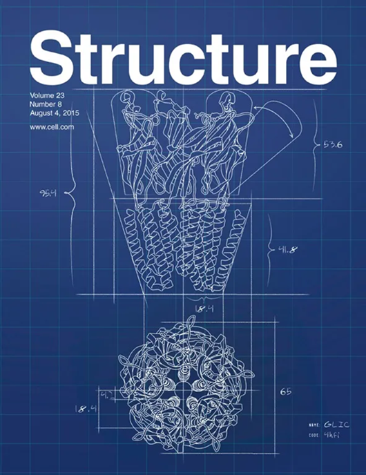
Structure
Structure 生物-生化与分子生物学
CiteScore
8.90
自引率
1.80%
发文量
155
审稿时长
3-8 weeks
期刊介绍: Structure aims to publish papers of exceptional interest in the field of structural biology. The journal strives to be essential reading for structural biologists, as well as biologists and biochemists that are interested in macromolecular structure and function. Structure strongly encourages the submission of manuscripts that present structural and molecular insights into biological function and mechanism. Other reports that address fundamental questions in structural biology, such as structure-based examinations of protein evolution, folding, and/or design, will also be considered. We will consider the application of any method, experimental or computational, at high or low resolution, to conduct structural investigations, as long as the method is appropriate for the biological, functional, and mechanistic question(s) being addressed. Likewise, reports describing single-molecule analysis of biological mechanisms are welcome. In general, the editors encourage submission of experimental structural studies that are enriched by an analysis of structure-activity relationships and will not consider studies that solely report structural information unless the structure or analysis is of exceptional and broad interest. Studies reporting only homology models, de novo models, or molecular dynamics simulations are also discouraged unless the models are informed by or validated by novel experimental data; rationalization of a large body of existing experimental evidence and making testable predictions based on a model or simulation is often not considered sufficient.
×
引用
GB/T 7714-2015
复制
MLA
复制
APA
复制
导出至
BibTeX EndNote RefMan NoteFirst NoteExpress
×
提示
您的信息不完整,为了账户安全,请先补充。
现在去补充
×
提示
您因"违规操作"
具体请查看互助需知
我知道了
×
提示
确定
请完成安全验证×
copy
已复制链接
快去分享给好友吧!
我知道了
右上角分享
点击右上角分享
0
联系我们:info@booksci.cn Book学术提供免费学术资源搜索服务,方便国内外学者检索中英文文献。致力于提供最便捷和优质的服务体验。 Copyright © 2023 布克学术 All rights reserved.
京ICP备2023020795号-1
ghs 京公网安备 11010802042870号
Book学术文献互助
Book学术文献互助群
群 号:481959085
Book学术官方微信