Roman Peter, Petr Hrobar, Josef Navratil, Martin Vagenknecht, Jindrich Soukup, Keiko Tsuji, Nestor X Barrezueta, Anna C Stoll, Renee C Gentzel, Jonathan A Sugam, Jacob Marcus, Danny A Bitton
{"title":"AnNoBrainer,利用深度学习自动标注小鼠大脑图像。","authors":"Roman Peter, Petr Hrobar, Josef Navratil, Martin Vagenknecht, Jindrich Soukup, Keiko Tsuji, Nestor X Barrezueta, Anna C Stoll, Renee C Gentzel, Jonathan A Sugam, Jacob Marcus, Danny A Bitton","doi":"10.1007/s12021-024-09679-1","DOIUrl":null,"url":null,"abstract":"<p><p>Annotation of multiple regions of interest across the whole mouse brain is an indispensable process for quantitative evaluation of a multitude of study endpoints in neuroscience digital pathology. Prior experience and domain expert knowledge are the key aspects for image annotation quality and consistency. At present, image annotation is often achieved manually by certified pathologists or trained technicians, limiting the total throughput of studies performed at neuroscience digital pathology labs. It may also mean that simpler and quicker methods of examining tissue samples are used by non-pathologists, especially in the early stages of research and preclinical studies. To address these limitations and to meet the growing demand for image analysis in a pharmaceutical setting, we developed AnNoBrainer, an open-source software tool that leverages deep learning, image registration, and standard cortical brain templates to automatically annotate individual brain regions on 2D pathology slides. Application of AnNoBrainer to a published set of pathology slides from transgenic mice models of synucleinopathy revealed comparable accuracy, increased reproducibility, and a significant reduction (~ 50%) in time spent on brain annotation, quality control and labelling compared to trained scientists in pathology. Taken together, AnNoBrainer offers a rapid, accurate, and reproducible automated annotation of mouse brain images that largely meets the experts' histopathological assessment standards (> 85% of cases) and enables high-throughput image analysis workflows in digital pathology labs.</p>","PeriodicalId":49761,"journal":{"name":"Neuroinformatics","volume":" ","pages":"719-730"},"PeriodicalIF":3.1000,"publicationDate":"2024-10-01","publicationTypes":"Journal Article","fieldsOfStudy":null,"isOpenAccess":false,"openAccessPdf":"https://www.ncbi.nlm.nih.gov/pmc/articles/PMC11579091/pdf/","citationCount":"0","resultStr":"{\"title\":\"AnNoBrainer, An Automated Annotation of Mouse Brain Images using Deep Learning.\",\"authors\":\"Roman Peter, Petr Hrobar, Josef Navratil, Martin Vagenknecht, Jindrich Soukup, Keiko Tsuji, Nestor X Barrezueta, Anna C Stoll, Renee C Gentzel, Jonathan A Sugam, Jacob Marcus, Danny A Bitton\",\"doi\":\"10.1007/s12021-024-09679-1\",\"DOIUrl\":null,\"url\":null,\"abstract\":\"<p><p>Annotation of multiple regions of interest across the whole mouse brain is an indispensable process for quantitative evaluation of a multitude of study endpoints in neuroscience digital pathology. Prior experience and domain expert knowledge are the key aspects for image annotation quality and consistency. At present, image annotation is often achieved manually by certified pathologists or trained technicians, limiting the total throughput of studies performed at neuroscience digital pathology labs. It may also mean that simpler and quicker methods of examining tissue samples are used by non-pathologists, especially in the early stages of research and preclinical studies. To address these limitations and to meet the growing demand for image analysis in a pharmaceutical setting, we developed AnNoBrainer, an open-source software tool that leverages deep learning, image registration, and standard cortical brain templates to automatically annotate individual brain regions on 2D pathology slides. Application of AnNoBrainer to a published set of pathology slides from transgenic mice models of synucleinopathy revealed comparable accuracy, increased reproducibility, and a significant reduction (~ 50%) in time spent on brain annotation, quality control and labelling compared to trained scientists in pathology. Taken together, AnNoBrainer offers a rapid, accurate, and reproducible automated annotation of mouse brain images that largely meets the experts' histopathological assessment standards (> 85% of cases) and enables high-throughput image analysis workflows in digital pathology labs.</p>\",\"PeriodicalId\":49761,\"journal\":{\"name\":\"Neuroinformatics\",\"volume\":\" \",\"pages\":\"719-730\"},\"PeriodicalIF\":3.1000,\"publicationDate\":\"2024-10-01\",\"publicationTypes\":\"Journal Article\",\"fieldsOfStudy\":null,\"isOpenAccess\":false,\"openAccessPdf\":\"https://www.ncbi.nlm.nih.gov/pmc/articles/PMC11579091/pdf/\",\"citationCount\":\"0\",\"resultStr\":null,\"platform\":\"Semanticscholar\",\"paperid\":null,\"PeriodicalName\":\"Neuroinformatics\",\"FirstCategoryId\":\"3\",\"ListUrlMain\":\"https://doi.org/10.1007/s12021-024-09679-1\",\"RegionNum\":4,\"RegionCategory\":\"医学\",\"ArticlePicture\":[],\"TitleCN\":null,\"AbstractTextCN\":null,\"PMCID\":null,\"EPubDate\":\"2024/8/7 0:00:00\",\"PubModel\":\"Epub\",\"JCR\":\"Q2\",\"JCRName\":\"COMPUTER SCIENCE, INTERDISCIPLINARY APPLICATIONS\",\"Score\":null,\"Total\":0}","platform":"Semanticscholar","paperid":null,"PeriodicalName":"Neuroinformatics","FirstCategoryId":"3","ListUrlMain":"https://doi.org/10.1007/s12021-024-09679-1","RegionNum":4,"RegionCategory":"医学","ArticlePicture":[],"TitleCN":null,"AbstractTextCN":null,"PMCID":null,"EPubDate":"2024/8/7 0:00:00","PubModel":"Epub","JCR":"Q2","JCRName":"COMPUTER SCIENCE, INTERDISCIPLINARY APPLICATIONS","Score":null,"Total":0}
AnNoBrainer, An Automated Annotation of Mouse Brain Images using Deep Learning.
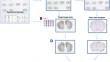
Annotation of multiple regions of interest across the whole mouse brain is an indispensable process for quantitative evaluation of a multitude of study endpoints in neuroscience digital pathology. Prior experience and domain expert knowledge are the key aspects for image annotation quality and consistency. At present, image annotation is often achieved manually by certified pathologists or trained technicians, limiting the total throughput of studies performed at neuroscience digital pathology labs. It may also mean that simpler and quicker methods of examining tissue samples are used by non-pathologists, especially in the early stages of research and preclinical studies. To address these limitations and to meet the growing demand for image analysis in a pharmaceutical setting, we developed AnNoBrainer, an open-source software tool that leverages deep learning, image registration, and standard cortical brain templates to automatically annotate individual brain regions on 2D pathology slides. Application of AnNoBrainer to a published set of pathology slides from transgenic mice models of synucleinopathy revealed comparable accuracy, increased reproducibility, and a significant reduction (~ 50%) in time spent on brain annotation, quality control and labelling compared to trained scientists in pathology. Taken together, AnNoBrainer offers a rapid, accurate, and reproducible automated annotation of mouse brain images that largely meets the experts' histopathological assessment standards (> 85% of cases) and enables high-throughput image analysis workflows in digital pathology labs.
期刊介绍:
Neuroinformatics publishes original articles and reviews with an emphasis on data structure and software tools related to analysis, modeling, integration, and sharing in all areas of neuroscience research. The editors particularly invite contributions on: (1) Theory and methodology, including discussions on ontologies, modeling approaches, database design, and meta-analyses; (2) Descriptions of developed databases and software tools, and of the methods for their distribution; (3) Relevant experimental results, such as reports accompanie by the release of massive data sets; (4) Computational simulations of models integrating and organizing complex data; and (5) Neuroengineering approaches, including hardware, robotics, and information theory studies.
 求助内容:
求助内容: 应助结果提醒方式:


