Ruoyu Shi, João Correia Pinto, Ivan Rienda, Peter Caie, Catarina Eloy, António Polónia
{"title":"明视野 HER2 原位杂交图像分析:临床应用验证。","authors":"Ruoyu Shi, João Correia Pinto, Ivan Rienda, Peter Caie, Catarina Eloy, António Polónia","doi":"10.1007/s00428-024-03889-3","DOIUrl":null,"url":null,"abstract":"<p><p>The aim of the present study was to develop and validate a quantitative image analysis (IA) algorithm to aid pathologists in assessing bright-field HER2 in situ hybridization (ISH) tests in solid cancers. A cohort of 80 sequential cases (40 HER2-negative and 40 HER2-positive) were evaluated for HER2 gene amplification with bright-field ISH. We developed an IA algorithm using the ISH Module from HALO software to automatically quantify HER2 and CEP17 copy numbers per cell as well as the HER2/CEP17 ratio. We observed a high correlation of HER2/CEP17 ratio, an average of HER2 and CEP17 copy number per cell between visual and IA quantification (Pearson's correlation coefficient of 0.842, 0.916, and 0.765, respectively). IA was able to count from 124 cells to 47,044 cells (median of 5565 cells). The margin of error for the visual quantification of the HER2/CEP17 ratio and of the average of HER2 copy number per cell decreased from a median of 0.23 to 0.02 and from a median of 0.49 to 0.04, respectively, in IA. Curve estimation regression models showed that a minimum of 469 or 953 invasive cancer cells per case is needed to reach an average margin of error below 0.1 for the HER2/CEP17 ratio or for the average of HER2 copy number per cell, respectively. Lastly, on average, a case took 212.1 s to execute the IA, which means that it evaluates about 130 cells/s and requires 6.7 s/mm<sup>2</sup>. The concordance of the IA software with the visual scoring was 95%, with a sensitivity of 90% and a specificity of 100%. All four discordant cases were able to achieve concordant results after the region of interest adjustment. In conclusion, this validation study underscores the usefulness of IA in HER2 ISH testing, displaying excellent concordance with visual scoring and significantly reducing margins of error.</p>","PeriodicalId":23514,"journal":{"name":"Virchows Archiv","volume":null,"pages":null},"PeriodicalIF":3.4000,"publicationDate":"2024-08-07","publicationTypes":"Journal Article","fieldsOfStudy":null,"isOpenAccess":false,"openAccessPdf":"","citationCount":"0","resultStr":"{\"title\":\"Image analysis for bright-field HER2 in situ hybridization: validation for clinical use.\",\"authors\":\"Ruoyu Shi, João Correia Pinto, Ivan Rienda, Peter Caie, Catarina Eloy, António Polónia\",\"doi\":\"10.1007/s00428-024-03889-3\",\"DOIUrl\":null,\"url\":null,\"abstract\":\"<p><p>The aim of the present study was to develop and validate a quantitative image analysis (IA) algorithm to aid pathologists in assessing bright-field HER2 in situ hybridization (ISH) tests in solid cancers. A cohort of 80 sequential cases (40 HER2-negative and 40 HER2-positive) were evaluated for HER2 gene amplification with bright-field ISH. We developed an IA algorithm using the ISH Module from HALO software to automatically quantify HER2 and CEP17 copy numbers per cell as well as the HER2/CEP17 ratio. We observed a high correlation of HER2/CEP17 ratio, an average of HER2 and CEP17 copy number per cell between visual and IA quantification (Pearson's correlation coefficient of 0.842, 0.916, and 0.765, respectively). IA was able to count from 124 cells to 47,044 cells (median of 5565 cells). The margin of error for the visual quantification of the HER2/CEP17 ratio and of the average of HER2 copy number per cell decreased from a median of 0.23 to 0.02 and from a median of 0.49 to 0.04, respectively, in IA. Curve estimation regression models showed that a minimum of 469 or 953 invasive cancer cells per case is needed to reach an average margin of error below 0.1 for the HER2/CEP17 ratio or for the average of HER2 copy number per cell, respectively. Lastly, on average, a case took 212.1 s to execute the IA, which means that it evaluates about 130 cells/s and requires 6.7 s/mm<sup>2</sup>. The concordance of the IA software with the visual scoring was 95%, with a sensitivity of 90% and a specificity of 100%. All four discordant cases were able to achieve concordant results after the region of interest adjustment. In conclusion, this validation study underscores the usefulness of IA in HER2 ISH testing, displaying excellent concordance with visual scoring and significantly reducing margins of error.</p>\",\"PeriodicalId\":23514,\"journal\":{\"name\":\"Virchows Archiv\",\"volume\":null,\"pages\":null},\"PeriodicalIF\":3.4000,\"publicationDate\":\"2024-08-07\",\"publicationTypes\":\"Journal Article\",\"fieldsOfStudy\":null,\"isOpenAccess\":false,\"openAccessPdf\":\"\",\"citationCount\":\"0\",\"resultStr\":null,\"platform\":\"Semanticscholar\",\"paperid\":null,\"PeriodicalName\":\"Virchows Archiv\",\"FirstCategoryId\":\"3\",\"ListUrlMain\":\"https://doi.org/10.1007/s00428-024-03889-3\",\"RegionNum\":3,\"RegionCategory\":\"医学\",\"ArticlePicture\":[],\"TitleCN\":null,\"AbstractTextCN\":null,\"PMCID\":null,\"EPubDate\":\"\",\"PubModel\":\"\",\"JCR\":\"Q1\",\"JCRName\":\"PATHOLOGY\",\"Score\":null,\"Total\":0}","platform":"Semanticscholar","paperid":null,"PeriodicalName":"Virchows Archiv","FirstCategoryId":"3","ListUrlMain":"https://doi.org/10.1007/s00428-024-03889-3","RegionNum":3,"RegionCategory":"医学","ArticlePicture":[],"TitleCN":null,"AbstractTextCN":null,"PMCID":null,"EPubDate":"","PubModel":"","JCR":"Q1","JCRName":"PATHOLOGY","Score":null,"Total":0}
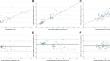
Image analysis for bright-field HER2 in situ hybridization: validation for clinical use.
The aim of the present study was to develop and validate a quantitative image analysis (IA) algorithm to aid pathologists in assessing bright-field HER2 in situ hybridization (ISH) tests in solid cancers. A cohort of 80 sequential cases (40 HER2-negative and 40 HER2-positive) were evaluated for HER2 gene amplification with bright-field ISH. We developed an IA algorithm using the ISH Module from HALO software to automatically quantify HER2 and CEP17 copy numbers per cell as well as the HER2/CEP17 ratio. We observed a high correlation of HER2/CEP17 ratio, an average of HER2 and CEP17 copy number per cell between visual and IA quantification (Pearson's correlation coefficient of 0.842, 0.916, and 0.765, respectively). IA was able to count from 124 cells to 47,044 cells (median of 5565 cells). The margin of error for the visual quantification of the HER2/CEP17 ratio and of the average of HER2 copy number per cell decreased from a median of 0.23 to 0.02 and from a median of 0.49 to 0.04, respectively, in IA. Curve estimation regression models showed that a minimum of 469 or 953 invasive cancer cells per case is needed to reach an average margin of error below 0.1 for the HER2/CEP17 ratio or for the average of HER2 copy number per cell, respectively. Lastly, on average, a case took 212.1 s to execute the IA, which means that it evaluates about 130 cells/s and requires 6.7 s/mm2. The concordance of the IA software with the visual scoring was 95%, with a sensitivity of 90% and a specificity of 100%. All four discordant cases were able to achieve concordant results after the region of interest adjustment. In conclusion, this validation study underscores the usefulness of IA in HER2 ISH testing, displaying excellent concordance with visual scoring and significantly reducing margins of error.
期刊介绍:
Manuscripts of original studies reinforcing the evidence base of modern diagnostic pathology, using immunocytochemical, molecular and ultrastructural techniques, will be welcomed. In addition, papers on critical evaluation of diagnostic criteria but also broadsheets and guidelines with a solid evidence base will be considered. Consideration will also be given to reports of work in other fields relevant to the understanding of human pathology as well as manuscripts on the application of new methods and techniques in pathology. Submission of purely experimental articles is discouraged but manuscripts on experimental work applicable to diagnostic pathology are welcomed. Biomarker studies are welcomed but need to abide by strict rules (e.g. REMARK) of adequate sample size and relevant marker choice. Single marker studies on limited patient series without validated application will as a rule not be considered. Case reports will only be considered when they provide substantial new information with an impact on understanding disease or diagnostic practice.

 求助内容:
求助内容: 应助结果提醒方式:


