{"title":"新一代测序技术在具有挑战性的肝脏 FNA 活检中的应用。","authors":"Dana J. Balitzer MD, Nancy Y. Greenland MD, PhD","doi":"10.1002/cncy.22893","DOIUrl":null,"url":null,"abstract":"<div>\n \n \n <section>\n \n <h3> Background</h3>\n \n <p>Fine-needle aspiration (FNA) biopsy is increasingly used for the diagnosis of hepatocellular masses. Because distinguishing well differentiated hepatocellular carcinoma (HCC) from other well differentiated hepatocellular lesions (e.g., large regenerative nodules or focal nodular hyperplasia) requires an assessment of architectural features, this may be challenging on FNA when intact tissue fragments are not sampled. Poorly differentiated HCC and intrahepatic cholangiocarcinoma (ICC) may exhibit overlapping pathologic features. Molecular testing can be helpful, because mutations in <i>TERT</i> promoter and <i>CTNNB1</i> (β-catenin) are characteristic of HCC, whereas mutations in <i>BAP1</i>, <i>IDH1/IDH2</i>, and <i>PBRM1</i> may favor ICC. The goal of this study was to assess the role of next-generation sequencing (NGS) in further subclassifying indeterminate liver lesions sampled by FNA.</p>\n </section>\n \n <section>\n \n <h3> Methods</h3>\n \n <p>A retrospective review of liver cytology cases with NGS on cell block material was performed. Age, radiologic features, background hepatic disease and treatment, outcome, and NGS data were obtained from the electronic medical record.</p>\n </section>\n \n <section>\n \n <h3> Results</h3>\n \n <p>Twelve FNA biopsies that had cell blocks from clinically suspected primary hepatic masses were identified. The presence of a <i>TERT</i> promoter mutation supported a diagnosis of HCC for one well differentiated neoplasm. For three patients, the presence of mutations, such as <i>IDH1</i>, <i>CDKN2A/CDKN2B</i>, and <i>BRAF</i>, supported a diagnosis of ICC. Of the eight poorly differentiated carcinomas, NGS helped refine the diagnosis in six of eight cases, with one HCC, three ICCs, and two that had combined HCC-ICC, with two cases remaining unclassified.</p>\n </section>\n \n <section>\n \n <h3> Conclusions</h3>\n \n <p>Molecular diagnostics can be helpful to distinguish HCC and ICC on FNA specimens, although a subset of primary hepatic tumors may remain unclassifiable.</p>\n </section>\n </div>","PeriodicalId":9410,"journal":{"name":"Cancer Cytopathology","volume":"132 11","pages":"714-722"},"PeriodicalIF":3.2000,"publicationDate":"2024-08-04","publicationTypes":"Journal Article","fieldsOfStudy":null,"isOpenAccess":false,"openAccessPdf":"https://onlinelibrary.wiley.com/doi/epdf/10.1002/cncy.22893","citationCount":"0","resultStr":"{\"title\":\"The utility of next-generation sequencing in challenging liver FNA biopsies\",\"authors\":\"Dana J. Balitzer MD, Nancy Y. Greenland MD, PhD\",\"doi\":\"10.1002/cncy.22893\",\"DOIUrl\":null,\"url\":null,\"abstract\":\"<div>\\n \\n \\n <section>\\n \\n <h3> Background</h3>\\n \\n <p>Fine-needle aspiration (FNA) biopsy is increasingly used for the diagnosis of hepatocellular masses. Because distinguishing well differentiated hepatocellular carcinoma (HCC) from other well differentiated hepatocellular lesions (e.g., large regenerative nodules or focal nodular hyperplasia) requires an assessment of architectural features, this may be challenging on FNA when intact tissue fragments are not sampled. Poorly differentiated HCC and intrahepatic cholangiocarcinoma (ICC) may exhibit overlapping pathologic features. Molecular testing can be helpful, because mutations in <i>TERT</i> promoter and <i>CTNNB1</i> (β-catenin) are characteristic of HCC, whereas mutations in <i>BAP1</i>, <i>IDH1/IDH2</i>, and <i>PBRM1</i> may favor ICC. The goal of this study was to assess the role of next-generation sequencing (NGS) in further subclassifying indeterminate liver lesions sampled by FNA.</p>\\n </section>\\n \\n <section>\\n \\n <h3> Methods</h3>\\n \\n <p>A retrospective review of liver cytology cases with NGS on cell block material was performed. Age, radiologic features, background hepatic disease and treatment, outcome, and NGS data were obtained from the electronic medical record.</p>\\n </section>\\n \\n <section>\\n \\n <h3> Results</h3>\\n \\n <p>Twelve FNA biopsies that had cell blocks from clinically suspected primary hepatic masses were identified. The presence of a <i>TERT</i> promoter mutation supported a diagnosis of HCC for one well differentiated neoplasm. For three patients, the presence of mutations, such as <i>IDH1</i>, <i>CDKN2A/CDKN2B</i>, and <i>BRAF</i>, supported a diagnosis of ICC. Of the eight poorly differentiated carcinomas, NGS helped refine the diagnosis in six of eight cases, with one HCC, three ICCs, and two that had combined HCC-ICC, with two cases remaining unclassified.</p>\\n </section>\\n \\n <section>\\n \\n <h3> Conclusions</h3>\\n \\n <p>Molecular diagnostics can be helpful to distinguish HCC and ICC on FNA specimens, although a subset of primary hepatic tumors may remain unclassifiable.</p>\\n </section>\\n </div>\",\"PeriodicalId\":9410,\"journal\":{\"name\":\"Cancer Cytopathology\",\"volume\":\"132 11\",\"pages\":\"714-722\"},\"PeriodicalIF\":3.2000,\"publicationDate\":\"2024-08-04\",\"publicationTypes\":\"Journal Article\",\"fieldsOfStudy\":null,\"isOpenAccess\":false,\"openAccessPdf\":\"https://onlinelibrary.wiley.com/doi/epdf/10.1002/cncy.22893\",\"citationCount\":\"0\",\"resultStr\":null,\"platform\":\"Semanticscholar\",\"paperid\":null,\"PeriodicalName\":\"Cancer Cytopathology\",\"FirstCategoryId\":\"3\",\"ListUrlMain\":\"https://acsjournals.onlinelibrary.wiley.com/doi/10.1002/cncy.22893\",\"RegionNum\":3,\"RegionCategory\":\"医学\",\"ArticlePicture\":[],\"TitleCN\":null,\"AbstractTextCN\":null,\"PMCID\":null,\"EPubDate\":\"\",\"PubModel\":\"\",\"JCR\":\"Q3\",\"JCRName\":\"ONCOLOGY\",\"Score\":null,\"Total\":0}","platform":"Semanticscholar","paperid":null,"PeriodicalName":"Cancer Cytopathology","FirstCategoryId":"3","ListUrlMain":"https://acsjournals.onlinelibrary.wiley.com/doi/10.1002/cncy.22893","RegionNum":3,"RegionCategory":"医学","ArticlePicture":[],"TitleCN":null,"AbstractTextCN":null,"PMCID":null,"EPubDate":"","PubModel":"","JCR":"Q3","JCRName":"ONCOLOGY","Score":null,"Total":0}
The utility of next-generation sequencing in challenging liver FNA biopsies
Background
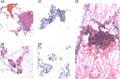
Fine-needle aspiration (FNA) biopsy is increasingly used for the diagnosis of hepatocellular masses. Because distinguishing well differentiated hepatocellular carcinoma (HCC) from other well differentiated hepatocellular lesions (e.g., large regenerative nodules or focal nodular hyperplasia) requires an assessment of architectural features, this may be challenging on FNA when intact tissue fragments are not sampled. Poorly differentiated HCC and intrahepatic cholangiocarcinoma (ICC) may exhibit overlapping pathologic features. Molecular testing can be helpful, because mutations in TERT promoter and CTNNB1 (β-catenin) are characteristic of HCC, whereas mutations in BAP1, IDH1/IDH2, and PBRM1 may favor ICC. The goal of this study was to assess the role of next-generation sequencing (NGS) in further subclassifying indeterminate liver lesions sampled by FNA.
Methods
A retrospective review of liver cytology cases with NGS on cell block material was performed. Age, radiologic features, background hepatic disease and treatment, outcome, and NGS data were obtained from the electronic medical record.
Results
Twelve FNA biopsies that had cell blocks from clinically suspected primary hepatic masses were identified. The presence of a TERT promoter mutation supported a diagnosis of HCC for one well differentiated neoplasm. For three patients, the presence of mutations, such as IDH1, CDKN2A/CDKN2B, and BRAF, supported a diagnosis of ICC. Of the eight poorly differentiated carcinomas, NGS helped refine the diagnosis in six of eight cases, with one HCC, three ICCs, and two that had combined HCC-ICC, with two cases remaining unclassified.
Conclusions
Molecular diagnostics can be helpful to distinguish HCC and ICC on FNA specimens, although a subset of primary hepatic tumors may remain unclassifiable.
期刊介绍:
Cancer Cytopathology provides a unique forum for interaction and dissemination of original research and educational information relevant to the practice of cytopathology and its related oncologic disciplines. The journal strives to have a positive effect on cancer prevention, early detection, diagnosis, and cure by the publication of high-quality content. The mission of Cancer Cytopathology is to present and inform readers of new applications, technological advances, cutting-edge research, novel applications of molecular techniques, and relevant review articles related to cytopathology.

 求助内容:
求助内容: 应助结果提醒方式:


