Fei Fei, Nivaz Brar, Melissa Beth Herring, Joshua R Menke, Jean Oak, Sebastian Fernandez-Pol
{"title":"外周血中真菌病/Sezary 综合征与非肿瘤性对照病例的 CD3 和 CD4 荧光强度中位数量化。","authors":"Fei Fei, Nivaz Brar, Melissa Beth Herring, Joshua R Menke, Jean Oak, Sebastian Fernandez-Pol","doi":"10.1007/s12308-024-00599-2","DOIUrl":null,"url":null,"abstract":"<p><p>Peripheral blood involvement by MF/SS has significant implications for prognosis and treatment. Flow cytometry is commonly used to assess MF/SS by analyzing the ratio of CD26- and/or CD7-CD4 + T cells and assessment of immunophenotypic abnormalities. However, distinguishing normal from abnormal cells is not always easy. In this study, we aimed to establish quantitative thresholds to better distinguish normal CD4 + T cells from neoplastic CD4 + T cells. A retrospective analysis of flow cytometry data was performed on 30 MF/SS patients with a detectable abnormal T cell population (positive), 63 patients with suspected or confirmed cutaneous involvement without a detectable abnormal T cell population (negative), and 60 healthy controls (control). CD3 and CD4 median fluorescence intensity (MFI) was normalized to internal control subsets. Among the positive cases, 50% had CD3 expression outside ± 2 SD from the mean of the negative and control group in the CD4 + CD26- subset. The corresponding specificity of this threshold was 94%. The ± 2 SD threshold showed a sensitivity of 57% and a specificity of 94% for the CD3 intensity among the CD7-negative subset. For CD4 intensity, the ± 2 SD threshold had a sensitivity of 33.3% and specificity of 95% for the CD26-negative subset and a sensitivity of 37% and specificity of 95% for the CD7-negative subset. In our study, although changes in CD3 and CD4 intensity greater than ± 2 SD were specific for MF/SS, more subtle differences in the intensity of CD3 and CD4 should not be used as the sole abnormality to make a diagnosis of circulating MF/SS.</p>","PeriodicalId":51320,"journal":{"name":"Journal of Hematopathology","volume":" ","pages":"191-199"},"PeriodicalIF":0.6000,"publicationDate":"2024-12-01","publicationTypes":"Journal Article","fieldsOfStudy":null,"isOpenAccess":false,"openAccessPdf":"","citationCount":"0","resultStr":"{\"title\":\"Quantification of the median fluorescence intensity of CD3 and CD4 in mycosis fungoides/Sezary syndrome versus non-neoplastic control cases in peripheral blood.\",\"authors\":\"Fei Fei, Nivaz Brar, Melissa Beth Herring, Joshua R Menke, Jean Oak, Sebastian Fernandez-Pol\",\"doi\":\"10.1007/s12308-024-00599-2\",\"DOIUrl\":null,\"url\":null,\"abstract\":\"<p><p>Peripheral blood involvement by MF/SS has significant implications for prognosis and treatment. Flow cytometry is commonly used to assess MF/SS by analyzing the ratio of CD26- and/or CD7-CD4 + T cells and assessment of immunophenotypic abnormalities. However, distinguishing normal from abnormal cells is not always easy. In this study, we aimed to establish quantitative thresholds to better distinguish normal CD4 + T cells from neoplastic CD4 + T cells. A retrospective analysis of flow cytometry data was performed on 30 MF/SS patients with a detectable abnormal T cell population (positive), 63 patients with suspected or confirmed cutaneous involvement without a detectable abnormal T cell population (negative), and 60 healthy controls (control). CD3 and CD4 median fluorescence intensity (MFI) was normalized to internal control subsets. Among the positive cases, 50% had CD3 expression outside ± 2 SD from the mean of the negative and control group in the CD4 + CD26- subset. The corresponding specificity of this threshold was 94%. The ± 2 SD threshold showed a sensitivity of 57% and a specificity of 94% for the CD3 intensity among the CD7-negative subset. For CD4 intensity, the ± 2 SD threshold had a sensitivity of 33.3% and specificity of 95% for the CD26-negative subset and a sensitivity of 37% and specificity of 95% for the CD7-negative subset. In our study, although changes in CD3 and CD4 intensity greater than ± 2 SD were specific for MF/SS, more subtle differences in the intensity of CD3 and CD4 should not be used as the sole abnormality to make a diagnosis of circulating MF/SS.</p>\",\"PeriodicalId\":51320,\"journal\":{\"name\":\"Journal of Hematopathology\",\"volume\":\" \",\"pages\":\"191-199\"},\"PeriodicalIF\":0.6000,\"publicationDate\":\"2024-12-01\",\"publicationTypes\":\"Journal Article\",\"fieldsOfStudy\":null,\"isOpenAccess\":false,\"openAccessPdf\":\"\",\"citationCount\":\"0\",\"resultStr\":null,\"platform\":\"Semanticscholar\",\"paperid\":null,\"PeriodicalName\":\"Journal of Hematopathology\",\"FirstCategoryId\":\"3\",\"ListUrlMain\":\"https://doi.org/10.1007/s12308-024-00599-2\",\"RegionNum\":4,\"RegionCategory\":\"医学\",\"ArticlePicture\":[],\"TitleCN\":null,\"AbstractTextCN\":null,\"PMCID\":null,\"EPubDate\":\"2024/8/2 0:00:00\",\"PubModel\":\"Epub\",\"JCR\":\"Q4\",\"JCRName\":\"HEMATOLOGY\",\"Score\":null,\"Total\":0}","platform":"Semanticscholar","paperid":null,"PeriodicalName":"Journal of Hematopathology","FirstCategoryId":"3","ListUrlMain":"https://doi.org/10.1007/s12308-024-00599-2","RegionNum":4,"RegionCategory":"医学","ArticlePicture":[],"TitleCN":null,"AbstractTextCN":null,"PMCID":null,"EPubDate":"2024/8/2 0:00:00","PubModel":"Epub","JCR":"Q4","JCRName":"HEMATOLOGY","Score":null,"Total":0}
Quantification of the median fluorescence intensity of CD3 and CD4 in mycosis fungoides/Sezary syndrome versus non-neoplastic control cases in peripheral blood.
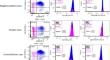
Peripheral blood involvement by MF/SS has significant implications for prognosis and treatment. Flow cytometry is commonly used to assess MF/SS by analyzing the ratio of CD26- and/or CD7-CD4 + T cells and assessment of immunophenotypic abnormalities. However, distinguishing normal from abnormal cells is not always easy. In this study, we aimed to establish quantitative thresholds to better distinguish normal CD4 + T cells from neoplastic CD4 + T cells. A retrospective analysis of flow cytometry data was performed on 30 MF/SS patients with a detectable abnormal T cell population (positive), 63 patients with suspected or confirmed cutaneous involvement without a detectable abnormal T cell population (negative), and 60 healthy controls (control). CD3 and CD4 median fluorescence intensity (MFI) was normalized to internal control subsets. Among the positive cases, 50% had CD3 expression outside ± 2 SD from the mean of the negative and control group in the CD4 + CD26- subset. The corresponding specificity of this threshold was 94%. The ± 2 SD threshold showed a sensitivity of 57% and a specificity of 94% for the CD3 intensity among the CD7-negative subset. For CD4 intensity, the ± 2 SD threshold had a sensitivity of 33.3% and specificity of 95% for the CD26-negative subset and a sensitivity of 37% and specificity of 95% for the CD7-negative subset. In our study, although changes in CD3 and CD4 intensity greater than ± 2 SD were specific for MF/SS, more subtle differences in the intensity of CD3 and CD4 should not be used as the sole abnormality to make a diagnosis of circulating MF/SS.
期刊介绍:
The Journal of Hematopathology aims at providing pathologists with a special interest in hematopathology with all the information needed to perform modern pathology in evaluating lymphoid tissues and bone marrow. To this end the journal publishes reviews, editorials, comments, original papers, guidelines and protocols, papers on ancillary techniques, and occasional case reports in the fields of the pathology, molecular biology, and clinical features of diseases of the hematopoietic system.
The journal is the unique reference point for all pathologists with an interest in hematopathology. Molecular biologists involved in the expanding field of molecular diagnostics and research on lymphomas and leukemia benefit from the journal, too. Furthermore, the journal is of major interest for hematologists dealing with patients suffering from lymphomas, leukemias, and other diseases.
The journal is unique in its true international character. Especially in the field of hematopathology it is clear that there are huge geographical variations in incidence of diseases. This is not only locally relevant, but due to globalization, relevant for all those involved in the management of patients.
 求助内容:
求助内容: 应助结果提醒方式:


