{"title":"非缺血性视网膜中央静脉闭塞患者视网膜各层厚度的纵向变化。","authors":"Min-Woo Lee, Ji-Ho Jun, Hyun-Je Seong","doi":"10.1186/s40662-024-00397-y","DOIUrl":null,"url":null,"abstract":"<p><strong>Background: </strong>To identify longitudinal changes in each retinal layer thickness in central retinal vein occlusion (CRVO) patients with resolved macular edema (ME).</p><p><strong>Methods: </strong>In this retrospective observational study, CRVO patients without a recurrence of ME for more than 3 years and normal controls were enrolled. Each retinal layer thickness of the parafoveal area, including ganglion cell complex (GCC), inner nuclear layer (INL), outer plexiform layer (OPL), outer nuclear layer (ONL), photoreceptor layer (PRL), and retinal pigment epithelium (RPE) was measured. After the resolution of ME, three more examinations with a 1-year interval were analyzed.</p><p><strong>Results: </strong>A total of 98 eyes were enrolled, 50 eyes for the control group and 48 eyes for the CRVO group. The baseline GCC thickness was 114.2 ± 15.6 μm and 104.2 ± 25.4 μm in the control and CRVO groups, respectively, which was significantly different (P = 0.022). The thicknesses of other layers including INL, OPL, ONL, PRL, and RPE were not significantly different at baseline. The reduction rate of GCC, INL, OPL, and ONL was - 3.92, - 1.33, - 0.91, and - 2.31 μm/year in the CRVO group, whereas no significant reductions were observed in the control group. Best-corrected visual acuity was significantly associated with changes in the GCC, OPL, and ONL in the CRVO group.</p><p><strong>Conclusions: </strong>In patients with CRVO, even in the absence of recurrent ME, retinal damage progresses over time, evidenced by thinning of the inner retina and outer retina including OPL and ONL. These changes may be associated with alterations in visual function.</p>","PeriodicalId":12194,"journal":{"name":"Eye and Vision","volume":"11 1","pages":"29"},"PeriodicalIF":4.0000,"publicationDate":"2024-08-01","publicationTypes":"Journal Article","fieldsOfStudy":null,"isOpenAccess":false,"openAccessPdf":"https://www.ncbi.nlm.nih.gov/pmc/articles/PMC11293173/pdf/","citationCount":"0","resultStr":"{\"title\":\"Longitudinal changes in each retinal layer thickness in patients with non-ischemic central retinal vein occlusion.\",\"authors\":\"Min-Woo Lee, Ji-Ho Jun, Hyun-Je Seong\",\"doi\":\"10.1186/s40662-024-00397-y\",\"DOIUrl\":null,\"url\":null,\"abstract\":\"<p><strong>Background: </strong>To identify longitudinal changes in each retinal layer thickness in central retinal vein occlusion (CRVO) patients with resolved macular edema (ME).</p><p><strong>Methods: </strong>In this retrospective observational study, CRVO patients without a recurrence of ME for more than 3 years and normal controls were enrolled. Each retinal layer thickness of the parafoveal area, including ganglion cell complex (GCC), inner nuclear layer (INL), outer plexiform layer (OPL), outer nuclear layer (ONL), photoreceptor layer (PRL), and retinal pigment epithelium (RPE) was measured. After the resolution of ME, three more examinations with a 1-year interval were analyzed.</p><p><strong>Results: </strong>A total of 98 eyes were enrolled, 50 eyes for the control group and 48 eyes for the CRVO group. The baseline GCC thickness was 114.2 ± 15.6 μm and 104.2 ± 25.4 μm in the control and CRVO groups, respectively, which was significantly different (P = 0.022). The thicknesses of other layers including INL, OPL, ONL, PRL, and RPE were not significantly different at baseline. The reduction rate of GCC, INL, OPL, and ONL was - 3.92, - 1.33, - 0.91, and - 2.31 μm/year in the CRVO group, whereas no significant reductions were observed in the control group. Best-corrected visual acuity was significantly associated with changes in the GCC, OPL, and ONL in the CRVO group.</p><p><strong>Conclusions: </strong>In patients with CRVO, even in the absence of recurrent ME, retinal damage progresses over time, evidenced by thinning of the inner retina and outer retina including OPL and ONL. These changes may be associated with alterations in visual function.</p>\",\"PeriodicalId\":12194,\"journal\":{\"name\":\"Eye and Vision\",\"volume\":\"11 1\",\"pages\":\"29\"},\"PeriodicalIF\":4.0000,\"publicationDate\":\"2024-08-01\",\"publicationTypes\":\"Journal Article\",\"fieldsOfStudy\":null,\"isOpenAccess\":false,\"openAccessPdf\":\"https://www.ncbi.nlm.nih.gov/pmc/articles/PMC11293173/pdf/\",\"citationCount\":\"0\",\"resultStr\":null,\"platform\":\"Semanticscholar\",\"paperid\":null,\"PeriodicalName\":\"Eye and Vision\",\"FirstCategoryId\":\"3\",\"ListUrlMain\":\"https://doi.org/10.1186/s40662-024-00397-y\",\"RegionNum\":1,\"RegionCategory\":\"医学\",\"ArticlePicture\":[],\"TitleCN\":null,\"AbstractTextCN\":null,\"PMCID\":null,\"EPubDate\":\"\",\"PubModel\":\"\",\"JCR\":\"Q1\",\"JCRName\":\"OPHTHALMOLOGY\",\"Score\":null,\"Total\":0}","platform":"Semanticscholar","paperid":null,"PeriodicalName":"Eye and Vision","FirstCategoryId":"3","ListUrlMain":"https://doi.org/10.1186/s40662-024-00397-y","RegionNum":1,"RegionCategory":"医学","ArticlePicture":[],"TitleCN":null,"AbstractTextCN":null,"PMCID":null,"EPubDate":"","PubModel":"","JCR":"Q1","JCRName":"OPHTHALMOLOGY","Score":null,"Total":0}
Longitudinal changes in each retinal layer thickness in patients with non-ischemic central retinal vein occlusion.
Background: To identify longitudinal changes in each retinal layer thickness in central retinal vein occlusion (CRVO) patients with resolved macular edema (ME).
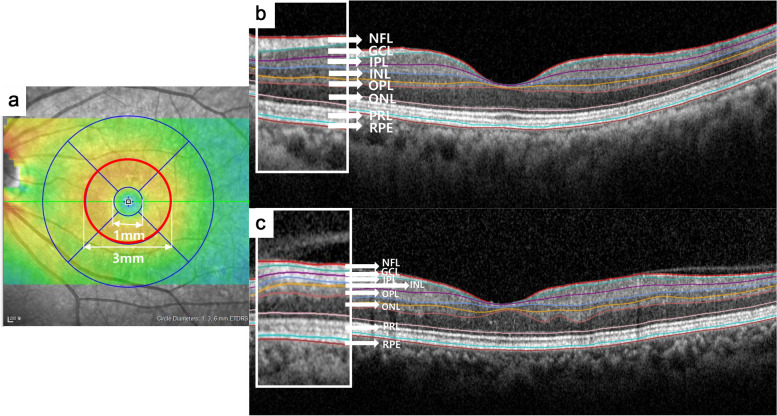
Methods: In this retrospective observational study, CRVO patients without a recurrence of ME for more than 3 years and normal controls were enrolled. Each retinal layer thickness of the parafoveal area, including ganglion cell complex (GCC), inner nuclear layer (INL), outer plexiform layer (OPL), outer nuclear layer (ONL), photoreceptor layer (PRL), and retinal pigment epithelium (RPE) was measured. After the resolution of ME, three more examinations with a 1-year interval were analyzed.
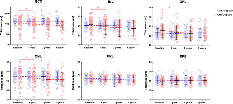
Results: A total of 98 eyes were enrolled, 50 eyes for the control group and 48 eyes for the CRVO group. The baseline GCC thickness was 114.2 ± 15.6 μm and 104.2 ± 25.4 μm in the control and CRVO groups, respectively, which was significantly different (P = 0.022). The thicknesses of other layers including INL, OPL, ONL, PRL, and RPE were not significantly different at baseline. The reduction rate of GCC, INL, OPL, and ONL was - 3.92, - 1.33, - 0.91, and - 2.31 μm/year in the CRVO group, whereas no significant reductions were observed in the control group. Best-corrected visual acuity was significantly associated with changes in the GCC, OPL, and ONL in the CRVO group.
Conclusions: In patients with CRVO, even in the absence of recurrent ME, retinal damage progresses over time, evidenced by thinning of the inner retina and outer retina including OPL and ONL. These changes may be associated with alterations in visual function.
期刊介绍:
Eye and Vision is an open access, peer-reviewed journal for ophthalmologists and visual science specialists. It welcomes research articles, reviews, methodologies, commentaries, case reports, perspectives and short reports encompassing all aspects of eye and vision. Topics of interest include but are not limited to: current developments of theoretical, experimental and clinical investigations in ophthalmology, optometry and vision science which focus on novel and high-impact findings on central issues pertaining to biology, pathophysiology and etiology of eye diseases as well as advances in diagnostic techniques, surgical treatment, instrument updates, the latest drug findings, results of clinical trials and research findings. It aims to provide ophthalmologists and visual science specialists with the latest developments in theoretical, experimental and clinical investigations in eye and vision.

 求助内容:
求助内容: 应助结果提醒方式:


