{"title":"利用高分辨率 3D 打印技术进行体外液芯纤维光度测量","authors":"Yu Chang , Can Wang , Ke Du","doi":"10.1016/j.snr.2024.100227","DOIUrl":null,"url":null,"abstract":"<div><p>High resolution 3D printing emerges as an alternative to microfabrication due to its fine resolution along with one-step manufacturing. Thus, it is broadly used in many fields, such as biological and chemical applications. We introduce such a technique to the design of the optofluidic probe by integrating optics and microfluidics as an ex vivo liquid core fiber photometry. We build the optofluidic probes with various T-shapes and conduct the transmission measurements and the ray tracing simulations, where the results are comparable. Through the transmission and fluorescence measurements, we obtain optimized curl T-shape dimensions of 524 µm wide, ∼50 µm thick, and 350 µm long with longitudinal spaces between them of 260 um. Furthermore, a heightened level of complexity in structure, characterized by a feature size of 25 µm, is attained through the improvement process. We conclude the feasibility of this optofluidic system with two applications: the in vivo-like setting consisting of thyroid biopsy training phantom and human plasma and the ex vivo-like setting consisting of the mice brain slices stained with wheat germ agglutinin (WGA). This prototype is an important step of establishing a 3D printing optofluidic applications for various in vivo research.</p></div>","PeriodicalId":426,"journal":{"name":"Sensors and Actuators Reports","volume":"8 ","pages":"Article 100227"},"PeriodicalIF":6.5000,"publicationDate":"2024-07-20","publicationTypes":"Journal Article","fieldsOfStudy":null,"isOpenAccess":false,"openAccessPdf":"https://www.sciencedirect.com/science/article/pii/S2666053924000432/pdfft?md5=7ddbc188d7737ca52f35c36891b6c873&pid=1-s2.0-S2666053924000432-main.pdf","citationCount":"0","resultStr":"{\"title\":\"Ex vivo liquid core fiber photometry with high-resolution 3D printing\",\"authors\":\"Yu Chang , Can Wang , Ke Du\",\"doi\":\"10.1016/j.snr.2024.100227\",\"DOIUrl\":null,\"url\":null,\"abstract\":\"<div><p>High resolution 3D printing emerges as an alternative to microfabrication due to its fine resolution along with one-step manufacturing. Thus, it is broadly used in many fields, such as biological and chemical applications. We introduce such a technique to the design of the optofluidic probe by integrating optics and microfluidics as an ex vivo liquid core fiber photometry. We build the optofluidic probes with various T-shapes and conduct the transmission measurements and the ray tracing simulations, where the results are comparable. Through the transmission and fluorescence measurements, we obtain optimized curl T-shape dimensions of 524 µm wide, ∼50 µm thick, and 350 µm long with longitudinal spaces between them of 260 um. Furthermore, a heightened level of complexity in structure, characterized by a feature size of 25 µm, is attained through the improvement process. We conclude the feasibility of this optofluidic system with two applications: the in vivo-like setting consisting of thyroid biopsy training phantom and human plasma and the ex vivo-like setting consisting of the mice brain slices stained with wheat germ agglutinin (WGA). This prototype is an important step of establishing a 3D printing optofluidic applications for various in vivo research.</p></div>\",\"PeriodicalId\":426,\"journal\":{\"name\":\"Sensors and Actuators Reports\",\"volume\":\"8 \",\"pages\":\"Article 100227\"},\"PeriodicalIF\":6.5000,\"publicationDate\":\"2024-07-20\",\"publicationTypes\":\"Journal Article\",\"fieldsOfStudy\":null,\"isOpenAccess\":false,\"openAccessPdf\":\"https://www.sciencedirect.com/science/article/pii/S2666053924000432/pdfft?md5=7ddbc188d7737ca52f35c36891b6c873&pid=1-s2.0-S2666053924000432-main.pdf\",\"citationCount\":\"0\",\"resultStr\":null,\"platform\":\"Semanticscholar\",\"paperid\":null,\"PeriodicalName\":\"Sensors and Actuators Reports\",\"FirstCategoryId\":\"1085\",\"ListUrlMain\":\"https://www.sciencedirect.com/science/article/pii/S2666053924000432\",\"RegionNum\":0,\"RegionCategory\":null,\"ArticlePicture\":[],\"TitleCN\":null,\"AbstractTextCN\":null,\"PMCID\":null,\"EPubDate\":\"\",\"PubModel\":\"\",\"JCR\":\"Q1\",\"JCRName\":\"BIOTECHNOLOGY & APPLIED MICROBIOLOGY\",\"Score\":null,\"Total\":0}","platform":"Semanticscholar","paperid":null,"PeriodicalName":"Sensors and Actuators Reports","FirstCategoryId":"1085","ListUrlMain":"https://www.sciencedirect.com/science/article/pii/S2666053924000432","RegionNum":0,"RegionCategory":null,"ArticlePicture":[],"TitleCN":null,"AbstractTextCN":null,"PMCID":null,"EPubDate":"","PubModel":"","JCR":"Q1","JCRName":"BIOTECHNOLOGY & APPLIED MICROBIOLOGY","Score":null,"Total":0}
引用次数: 0
摘要
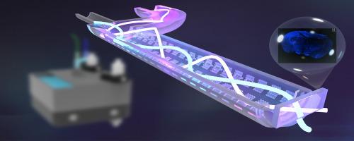
高分辨率三维打印因其精细的分辨率和一步法制造而成为微细加工的替代技术。因此,它被广泛应用于生物和化学等许多领域。我们将这种技术引入光流体探针的设计中,将光学和微流体学整合为体内外液芯光纤光度计。我们制作了不同 T 形的光流体探针,并进行了透射测量和光线追踪模拟,结果具有可比性。通过透射和荧光测量,我们获得了优化的卷曲 T 形尺寸:宽 524 微米、厚 50 微米、长 350 微米,它们之间的纵向空间为 260 微米。此外,通过改进工艺,结构的复杂程度也得到了提高,特征尺寸达到了 25 微米。我们通过两种应用总结了这一光流体系统的可行性:一种是由甲状腺活检训练模型和人体血浆组成的类活体环境,另一种是由小麦胚芽凝集素(WGA)染色的小鼠脑片组成的类活体环境。该原型是为各种体内研究建立 3D 打印光流体应用的重要一步。
Ex vivo liquid core fiber photometry with high-resolution 3D printing
High resolution 3D printing emerges as an alternative to microfabrication due to its fine resolution along with one-step manufacturing. Thus, it is broadly used in many fields, such as biological and chemical applications. We introduce such a technique to the design of the optofluidic probe by integrating optics and microfluidics as an ex vivo liquid core fiber photometry. We build the optofluidic probes with various T-shapes and conduct the transmission measurements and the ray tracing simulations, where the results are comparable. Through the transmission and fluorescence measurements, we obtain optimized curl T-shape dimensions of 524 µm wide, ∼50 µm thick, and 350 µm long with longitudinal spaces between them of 260 um. Furthermore, a heightened level of complexity in structure, characterized by a feature size of 25 µm, is attained through the improvement process. We conclude the feasibility of this optofluidic system with two applications: the in vivo-like setting consisting of thyroid biopsy training phantom and human plasma and the ex vivo-like setting consisting of the mice brain slices stained with wheat germ agglutinin (WGA). This prototype is an important step of establishing a 3D printing optofluidic applications for various in vivo research.
期刊介绍:
Sensors and Actuators Reports is a peer-reviewed open access journal launched out from the Sensors and Actuators journal family. Sensors and Actuators Reports is dedicated to publishing new and original works in the field of all type of sensors and actuators, including bio-, chemical-, physical-, and nano- sensors and actuators, which demonstrates significant progress beyond the current state of the art. The journal regularly publishes original research papers, reviews, and short communications.
For research papers and short communications, the journal aims to publish the new and original work supported by experimental results and as such purely theoretical works are not accepted.
 求助内容:
求助内容: 应助结果提醒方式:


