{"title":"大鼠卵巢和子宫中毛细血管细胞的分布和形态特征:超微结构和免疫组化分析的启示。","authors":"Merjem Purelku, Hakan Sahin, Gozde Erkanli Senturk, Gamze Tanriverdi","doi":"10.1007/s00418-024-02313-w","DOIUrl":null,"url":null,"abstract":"<p><p>Telocytes (TCs) are characterized by a small oval-shaped cell body with long prolongations that are called telopods (Tps). PDGFR-β and c-kit markers may assist for the immunohistochemical identification of TCs; however, by these means they cannot be identified with absolute specificity. Transmission electron microscopy (TEM) is considered as a gold standard method for TCs observation. Studies on TCs in the female reproductive system are limited, and there is a lack of awareness regarding TCs in rat ovaries. We aimed to demonstrate the existence and morphology of TCs in rat ovaries, alongside previously studied TCs in rat uteri. Thus, ovaries and uteri from young adult Sprague-Dawley female rats (n = 8) with regular estrous cycles were collected. Then, left ovaries and uteri were proccessed for TEM analysis, while the right ones were used for immunohistochemistry. As a result, TCs were seen throughout the rat's ovarian stroma with their characteristic cell bodies, Tps, podomes (Pds) and podomers (Pdms). Tps were situated within the thecal layer of the follicles, surrounding the corpus luteum and blood vessels. Ovarian TCs were recognized to have relationship with other TCs/stromal cells. Subsequently, TCs were seen in stroma of endometrium with surrounding blood vessels and uterine glands, myometrium and perimetrium in rat uteri. There was also no statistical significance between the number of c-kit+ and PDGFR-β+ telocyte-like cells in both rat ovarian (p = 0.137) and endometrial stroma (p = 0.450). Further investigation of the roles and functions of TCs in the female reproductive system is needed.</p>","PeriodicalId":13107,"journal":{"name":"Histochemistry and Cell Biology","volume":" ","pages":"373-384"},"PeriodicalIF":2.1000,"publicationDate":"2024-11-01","publicationTypes":"Journal Article","fieldsOfStudy":null,"isOpenAccess":false,"openAccessPdf":"https://www.ncbi.nlm.nih.gov/pmc/articles/PMC11393091/pdf/","citationCount":"0","resultStr":"{\"title\":\"Distribution and morphologic characterization of telocytes in rat ovary and uterus: insights from ultrastructural and immunohistochemical analysis.\",\"authors\":\"Merjem Purelku, Hakan Sahin, Gozde Erkanli Senturk, Gamze Tanriverdi\",\"doi\":\"10.1007/s00418-024-02313-w\",\"DOIUrl\":null,\"url\":null,\"abstract\":\"<p><p>Telocytes (TCs) are characterized by a small oval-shaped cell body with long prolongations that are called telopods (Tps). PDGFR-β and c-kit markers may assist for the immunohistochemical identification of TCs; however, by these means they cannot be identified with absolute specificity. Transmission electron microscopy (TEM) is considered as a gold standard method for TCs observation. Studies on TCs in the female reproductive system are limited, and there is a lack of awareness regarding TCs in rat ovaries. We aimed to demonstrate the existence and morphology of TCs in rat ovaries, alongside previously studied TCs in rat uteri. Thus, ovaries and uteri from young adult Sprague-Dawley female rats (n = 8) with regular estrous cycles were collected. Then, left ovaries and uteri were proccessed for TEM analysis, while the right ones were used for immunohistochemistry. As a result, TCs were seen throughout the rat's ovarian stroma with their characteristic cell bodies, Tps, podomes (Pds) and podomers (Pdms). Tps were situated within the thecal layer of the follicles, surrounding the corpus luteum and blood vessels. Ovarian TCs were recognized to have relationship with other TCs/stromal cells. Subsequently, TCs were seen in stroma of endometrium with surrounding blood vessels and uterine glands, myometrium and perimetrium in rat uteri. There was also no statistical significance between the number of c-kit+ and PDGFR-β+ telocyte-like cells in both rat ovarian (p = 0.137) and endometrial stroma (p = 0.450). Further investigation of the roles and functions of TCs in the female reproductive system is needed.</p>\",\"PeriodicalId\":13107,\"journal\":{\"name\":\"Histochemistry and Cell Biology\",\"volume\":\" \",\"pages\":\"373-384\"},\"PeriodicalIF\":2.1000,\"publicationDate\":\"2024-11-01\",\"publicationTypes\":\"Journal Article\",\"fieldsOfStudy\":null,\"isOpenAccess\":false,\"openAccessPdf\":\"https://www.ncbi.nlm.nih.gov/pmc/articles/PMC11393091/pdf/\",\"citationCount\":\"0\",\"resultStr\":null,\"platform\":\"Semanticscholar\",\"paperid\":null,\"PeriodicalName\":\"Histochemistry and Cell Biology\",\"FirstCategoryId\":\"99\",\"ListUrlMain\":\"https://doi.org/10.1007/s00418-024-02313-w\",\"RegionNum\":4,\"RegionCategory\":\"生物学\",\"ArticlePicture\":[],\"TitleCN\":null,\"AbstractTextCN\":null,\"PMCID\":null,\"EPubDate\":\"2024/7/30 0:00:00\",\"PubModel\":\"Epub\",\"JCR\":\"Q4\",\"JCRName\":\"CELL BIOLOGY\",\"Score\":null,\"Total\":0}","platform":"Semanticscholar","paperid":null,"PeriodicalName":"Histochemistry and Cell Biology","FirstCategoryId":"99","ListUrlMain":"https://doi.org/10.1007/s00418-024-02313-w","RegionNum":4,"RegionCategory":"生物学","ArticlePicture":[],"TitleCN":null,"AbstractTextCN":null,"PMCID":null,"EPubDate":"2024/7/30 0:00:00","PubModel":"Epub","JCR":"Q4","JCRName":"CELL BIOLOGY","Score":null,"Total":0}
Distribution and morphologic characterization of telocytes in rat ovary and uterus: insights from ultrastructural and immunohistochemical analysis.
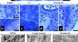
Telocytes (TCs) are characterized by a small oval-shaped cell body with long prolongations that are called telopods (Tps). PDGFR-β and c-kit markers may assist for the immunohistochemical identification of TCs; however, by these means they cannot be identified with absolute specificity. Transmission electron microscopy (TEM) is considered as a gold standard method for TCs observation. Studies on TCs in the female reproductive system are limited, and there is a lack of awareness regarding TCs in rat ovaries. We aimed to demonstrate the existence and morphology of TCs in rat ovaries, alongside previously studied TCs in rat uteri. Thus, ovaries and uteri from young adult Sprague-Dawley female rats (n = 8) with regular estrous cycles were collected. Then, left ovaries and uteri were proccessed for TEM analysis, while the right ones were used for immunohistochemistry. As a result, TCs were seen throughout the rat's ovarian stroma with their characteristic cell bodies, Tps, podomes (Pds) and podomers (Pdms). Tps were situated within the thecal layer of the follicles, surrounding the corpus luteum and blood vessels. Ovarian TCs were recognized to have relationship with other TCs/stromal cells. Subsequently, TCs were seen in stroma of endometrium with surrounding blood vessels and uterine glands, myometrium and perimetrium in rat uteri. There was also no statistical significance between the number of c-kit+ and PDGFR-β+ telocyte-like cells in both rat ovarian (p = 0.137) and endometrial stroma (p = 0.450). Further investigation of the roles and functions of TCs in the female reproductive system is needed.
期刊介绍:
Histochemistry and Cell Biology is devoted to the field of molecular histology and cell biology, publishing original articles dealing with the localization and identification of molecular components, metabolic activities and cell biological aspects of cells and tissues. Coverage extends to the development, application, and/or evaluation of methods and probes that can be used in the entire area of histochemistry and cell biology.
 求助内容:
求助内容: 应助结果提醒方式:


