封面图片,第 72 卷第 9 期

IF 5.4 2区 医学 Q1 NEUROSCIENCES
Glia Pub Date : 2024-07-21 DOI:10.1002/glia.24409
Paul F. Cullen, William J. Gammerdinger, Shannan J. Ho Sui, Arpan Guha Mazumder, Daniel Sun
{"title":"封面图片,第 72 卷第 9 期","authors":"Paul F. Cullen,&nbsp;William J. Gammerdinger,&nbsp;Shannan J. Ho Sui,&nbsp;Arpan Guha Mazumder,&nbsp;Daniel Sun","doi":"10.1002/glia.24409","DOIUrl":null,"url":null,"abstract":"<p>Cover Illustration: Confocal image of retinal astrocyte (green) isolated by microdissection from GFAP-Cre+ x Rpl22<sup>HA/HA</sup> mouse showing HA-tagged ribosomes (red) present throughout the cell, including within endfeet processes, indicating that ribosome associated mRNA throughout the cell can be isolated by immunoprecipitation. The image also indicates how well astrocyte morphology are preserved in our microdissection method. Nuclei are labeled with DAPI in blue. (See Cullen, P., et al, https://doi.org/10.1002/glia.24571)\n\n <figure>\n <div><picture>\n <source></source></picture><p></p>\n </div>\n </figure>\n </p>","PeriodicalId":174,"journal":{"name":"Glia","volume":"72 9","pages":"C1"},"PeriodicalIF":5.4000,"publicationDate":"2024-07-21","publicationTypes":"Journal Article","fieldsOfStudy":null,"isOpenAccess":false,"openAccessPdf":"https://onlinelibrary.wiley.com/doi/epdf/10.1002/glia.24409","citationCount":"0","resultStr":"{\"title\":\"Cover Image, Volume 72, Issue 9\",\"authors\":\"Paul F. Cullen,&nbsp;William J. Gammerdinger,&nbsp;Shannan J. Ho Sui,&nbsp;Arpan Guha Mazumder,&nbsp;Daniel Sun\",\"doi\":\"10.1002/glia.24409\",\"DOIUrl\":null,\"url\":null,\"abstract\":\"<p>Cover Illustration: Confocal image of retinal astrocyte (green) isolated by microdissection from GFAP-Cre+ x Rpl22<sup>HA/HA</sup> mouse showing HA-tagged ribosomes (red) present throughout the cell, including within endfeet processes, indicating that ribosome associated mRNA throughout the cell can be isolated by immunoprecipitation. The image also indicates how well astrocyte morphology are preserved in our microdissection method. Nuclei are labeled with DAPI in blue. (See Cullen, P., et al, https://doi.org/10.1002/glia.24571)\\n\\n <figure>\\n <div><picture>\\n <source></source></picture><p></p>\\n </div>\\n </figure>\\n </p>\",\"PeriodicalId\":174,\"journal\":{\"name\":\"Glia\",\"volume\":\"72 9\",\"pages\":\"C1\"},\"PeriodicalIF\":5.4000,\"publicationDate\":\"2024-07-21\",\"publicationTypes\":\"Journal Article\",\"fieldsOfStudy\":null,\"isOpenAccess\":false,\"openAccessPdf\":\"https://onlinelibrary.wiley.com/doi/epdf/10.1002/glia.24409\",\"citationCount\":\"0\",\"resultStr\":null,\"platform\":\"Semanticscholar\",\"paperid\":null,\"PeriodicalName\":\"Glia\",\"FirstCategoryId\":\"3\",\"ListUrlMain\":\"https://onlinelibrary.wiley.com/doi/10.1002/glia.24409\",\"RegionNum\":2,\"RegionCategory\":\"医学\",\"ArticlePicture\":[],\"TitleCN\":null,\"AbstractTextCN\":null,\"PMCID\":null,\"EPubDate\":\"\",\"PubModel\":\"\",\"JCR\":\"Q1\",\"JCRName\":\"NEUROSCIENCES\",\"Score\":null,\"Total\":0}","platform":"Semanticscholar","paperid":null,"PeriodicalName":"Glia","FirstCategoryId":"3","ListUrlMain":"https://onlinelibrary.wiley.com/doi/10.1002/glia.24409","RegionNum":2,"RegionCategory":"医学","ArticlePicture":[],"TitleCN":null,"AbstractTextCN":null,"PMCID":null,"EPubDate":"","PubModel":"","JCR":"Q1","JCRName":"NEUROSCIENCES","Score":null,"Total":0}
引用次数: 0

摘要

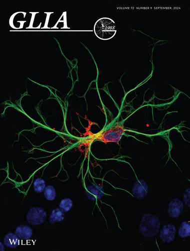
封面插图:通过显微切割从 GFAP-Cre+ x Rpl22HA/HA 小鼠体内分离出的视网膜星形胶质细胞(绿色)的共焦图像显示,HA 标记的核糖体(红色)遍布整个细胞,包括内膜过程,这表明通过免疫沉淀可以分离出整个细胞中与核糖体相关的 mRNA。该图像还显示了我们的显微切割方法对星形胶质细胞形态的良好保存。细胞核用蓝色 DAPI 标记。(见 Cullen, P. 等人,https://doi.org/10.1002/glia.24571)
本文章由计算机程序翻译,如有差异,请以英文原文为准。

Cover Image, Volume 72, Issue 9

Cover Image, Volume 72, Issue 9

Cover Illustration: Confocal image of retinal astrocyte (green) isolated by microdissection from GFAP-Cre+ x Rpl22HA/HA mouse showing HA-tagged ribosomes (red) present throughout the cell, including within endfeet processes, indicating that ribosome associated mRNA throughout the cell can be isolated by immunoprecipitation. The image also indicates how well astrocyte morphology are preserved in our microdissection method. Nuclei are labeled with DAPI in blue. (See Cullen, P., et al, https://doi.org/10.1002/glia.24571)

求助全文
通过发布文献求助,成功后即可免费获取论文全文。 去求助
来源期刊
Glia
Glia 医学-神经科学
CiteScore
13.10
自引率
4.80%
发文量
162
审稿时长
3-8 weeks
期刊介绍: GLIA is a peer-reviewed journal, which publishes articles dealing with all aspects of glial structure and function. This includes all aspects of glial cell biology in health and disease.
×
引用
GB/T 7714-2015
复制
MLA
复制
APA
复制
导出至
BibTeX EndNote RefMan NoteFirst NoteExpress
×
提示
您的信息不完整,为了账户安全,请先补充。
现在去补充
×
提示
您因"违规操作"
具体请查看互助需知
我知道了
×
提示
确定
请完成安全验证×
copy
已复制链接
快去分享给好友吧!
我知道了
右上角分享
点击右上角分享
0
联系我们:info@booksci.cn Book学术提供免费学术资源搜索服务,方便国内外学者检索中英文文献。致力于提供最便捷和优质的服务体验。 Copyright © 2023 布克学术 All rights reserved.
京ICP备2023020795号-1
ghs 京公网安备 11010802042870号
Book学术文献互助
Book学术文献互助群
群 号:481959085
Book学术官方微信