Sami A Al-Ani, Danny Chandla, John Delieu, Sinling Tiffany Yu, Antonio Fratini, Renia Gkountiou, Claire J Stocker
{"title":"使用三维足踝拼图可增强学生在医学院早期对骨骼解剖的理解。","authors":"Sami A Al-Ani, Danny Chandla, John Delieu, Sinling Tiffany Yu, Antonio Fratini, Renia Gkountiou, Claire J Stocker","doi":"10.1007/s00276-024-03439-1","DOIUrl":null,"url":null,"abstract":"<p><strong>Purpose: </strong>3D visualization is an important part of learning anatomy with cadavers generally used to effectuate this. However, high cost, ethical considerations, and limited accessibility can often limit the suitability of cadavers as teaching tools. Anatomical 3D printed models offer an alternative tool for teaching gross anatomy due to their low cost and accessibility. This study aims to investigate if combing gamification with 3D printed models can enhance the learning experience and be effective for teaching anatomy.</p><p><strong>Methods: </strong>3D printed models of the bones of the foot and ankle were generated, and 267 first-year medical students from 2 consecutive cohorts worked in groups to put it together as a puzzle. Participants completed a questionnaire regarding perceptions of 3D models and their knowledge of foot anatomy, before and after the session and were asked to provide comments.</p><p><strong>Results: </strong>Analysis of the responses showed a significant increase in the confidence of the learners in their anatomy knowledge and an increased appreciation of the role that 3D models have in enhancing the learning experience. After the session, there were many comments saying how enjoyable and engaging 3D models were.</p><p><strong>Conclusion: </strong>Through the puzzle element of the session, the students were challenged mentally to work out the anatomical features of the foot and ankle. The combined elements of the puzzle and the features of the 3D model assembly made the activity fun and conducive to active learning. The possibility of having fun was not something the students had considered before the session.</p>","PeriodicalId":49461,"journal":{"name":"Surgical and Radiologic Anatomy","volume":" ","pages":"1429-1438"},"PeriodicalIF":1.4000,"publicationDate":"2024-09-01","publicationTypes":"Journal Article","fieldsOfStudy":null,"isOpenAccess":false,"openAccessPdf":"https://www.ncbi.nlm.nih.gov/pmc/articles/PMC11322274/pdf/","citationCount":"0","resultStr":"{\"title\":\"Use of 3D foot and ankle puzzle enhances student understanding of the skeletal anatomy in the early years of medical school.\",\"authors\":\"Sami A Al-Ani, Danny Chandla, John Delieu, Sinling Tiffany Yu, Antonio Fratini, Renia Gkountiou, Claire J Stocker\",\"doi\":\"10.1007/s00276-024-03439-1\",\"DOIUrl\":null,\"url\":null,\"abstract\":\"<p><strong>Purpose: </strong>3D visualization is an important part of learning anatomy with cadavers generally used to effectuate this. However, high cost, ethical considerations, and limited accessibility can often limit the suitability of cadavers as teaching tools. Anatomical 3D printed models offer an alternative tool for teaching gross anatomy due to their low cost and accessibility. This study aims to investigate if combing gamification with 3D printed models can enhance the learning experience and be effective for teaching anatomy.</p><p><strong>Methods: </strong>3D printed models of the bones of the foot and ankle were generated, and 267 first-year medical students from 2 consecutive cohorts worked in groups to put it together as a puzzle. Participants completed a questionnaire regarding perceptions of 3D models and their knowledge of foot anatomy, before and after the session and were asked to provide comments.</p><p><strong>Results: </strong>Analysis of the responses showed a significant increase in the confidence of the learners in their anatomy knowledge and an increased appreciation of the role that 3D models have in enhancing the learning experience. After the session, there were many comments saying how enjoyable and engaging 3D models were.</p><p><strong>Conclusion: </strong>Through the puzzle element of the session, the students were challenged mentally to work out the anatomical features of the foot and ankle. The combined elements of the puzzle and the features of the 3D model assembly made the activity fun and conducive to active learning. The possibility of having fun was not something the students had considered before the session.</p>\",\"PeriodicalId\":49461,\"journal\":{\"name\":\"Surgical and Radiologic Anatomy\",\"volume\":\" \",\"pages\":\"1429-1438\"},\"PeriodicalIF\":1.4000,\"publicationDate\":\"2024-09-01\",\"publicationTypes\":\"Journal Article\",\"fieldsOfStudy\":null,\"isOpenAccess\":false,\"openAccessPdf\":\"https://www.ncbi.nlm.nih.gov/pmc/articles/PMC11322274/pdf/\",\"citationCount\":\"0\",\"resultStr\":null,\"platform\":\"Semanticscholar\",\"paperid\":null,\"PeriodicalName\":\"Surgical and Radiologic Anatomy\",\"FirstCategoryId\":\"3\",\"ListUrlMain\":\"https://doi.org/10.1007/s00276-024-03439-1\",\"RegionNum\":4,\"RegionCategory\":\"医学\",\"ArticlePicture\":[],\"TitleCN\":null,\"AbstractTextCN\":null,\"PMCID\":null,\"EPubDate\":\"2024/7/26 0:00:00\",\"PubModel\":\"Epub\",\"JCR\":\"Q2\",\"JCRName\":\"Medicine\",\"Score\":null,\"Total\":0}","platform":"Semanticscholar","paperid":null,"PeriodicalName":"Surgical and Radiologic Anatomy","FirstCategoryId":"3","ListUrlMain":"https://doi.org/10.1007/s00276-024-03439-1","RegionNum":4,"RegionCategory":"医学","ArticlePicture":[],"TitleCN":null,"AbstractTextCN":null,"PMCID":null,"EPubDate":"2024/7/26 0:00:00","PubModel":"Epub","JCR":"Q2","JCRName":"Medicine","Score":null,"Total":0}
引用次数: 0
摘要
目的:三维可视化是学习解剖学的一个重要部分,通常使用尸体来实现这一点。然而,高昂的成本、伦理方面的考虑以及有限的可及性往往限制了尸体作为教学工具的适用性。解剖学 3D 打印模型因其低成本和易获取性,为大体解剖学教学提供了另一种工具。本研究旨在探讨将游戏化与 3D 打印模型相结合是否能增强学习体验并有效地进行解剖学教学。方法:我们制作了足部和踝部骨骼的 3D 打印模型,来自连续两届的 267 名一年级医学生以小组为单位将其拼成一个拼图。参与者在课程前后填写了一份关于对 3D 模型的看法和足部解剖知识的问卷,并被要求提供意见:结果:对回答的分析表明,学员对解剖学知识的信心明显增强,对 3D 模型在增强学习体验方面的作用的认识也有所提高。课程结束后,许多人评论说三维模型是多么令人愉快和引人入胜:通过拼图环节,学生们在脑力上受到了挑战,要拼出足部和踝部的解剖特征。拼图元素与三维模型组装的特点相结合,使活动充满乐趣,有利于学生主动学习。在这节课之前,学生们并没有考虑过玩得开心的可能性。
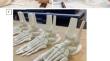
Use of 3D foot and ankle puzzle enhances student understanding of the skeletal anatomy in the early years of medical school.
Purpose: 3D visualization is an important part of learning anatomy with cadavers generally used to effectuate this. However, high cost, ethical considerations, and limited accessibility can often limit the suitability of cadavers as teaching tools. Anatomical 3D printed models offer an alternative tool for teaching gross anatomy due to their low cost and accessibility. This study aims to investigate if combing gamification with 3D printed models can enhance the learning experience and be effective for teaching anatomy.
Methods: 3D printed models of the bones of the foot and ankle were generated, and 267 first-year medical students from 2 consecutive cohorts worked in groups to put it together as a puzzle. Participants completed a questionnaire regarding perceptions of 3D models and their knowledge of foot anatomy, before and after the session and were asked to provide comments.
Results: Analysis of the responses showed a significant increase in the confidence of the learners in their anatomy knowledge and an increased appreciation of the role that 3D models have in enhancing the learning experience. After the session, there were many comments saying how enjoyable and engaging 3D models were.
Conclusion: Through the puzzle element of the session, the students were challenged mentally to work out the anatomical features of the foot and ankle. The combined elements of the puzzle and the features of the 3D model assembly made the activity fun and conducive to active learning. The possibility of having fun was not something the students had considered before the session.
期刊介绍:
Anatomy is a morphological science which cannot fail to interest the clinician. The practical application of anatomical research to clinical problems necessitates special adaptation and selectivity in choosing from numerous international works. Although there is a tendency to believe that meaningful advances in anatomy are unlikely, constant revision is necessary. Surgical and Radiologic Anatomy, the first international journal of Clinical anatomy has been created in this spirit.
Its goal is to serve clinicians, regardless of speciality-physicians, surgeons, radiologists or other specialists-as an indispensable aid with which they can improve their knowledge of anatomy. Each issue includes: Original papers, review articles, articles on the anatomical bases of medical, surgical and radiological techniques, articles of normal radiologic anatomy, brief reviews of anatomical publications of clinical interest.
Particular attention is given to high quality illustrations, which are indispensable for a better understanding of anatomical problems.
Surgical and Radiologic Anatomy is a journal written by anatomists for clinicians with a special interest in anatomy.

 求助内容:
求助内容: 应助结果提醒方式:


