Jin Li, Junxia Niu, Weimin Zheng, Yueyan Bian, Fang Wu, Xiuqin Jia, Zhaoyang Fan, Xihai Zhao, Qi Yang
{"title":"全脑血管壁成像中扩张的皮质下动脉可区分发病机制并预测单发皮质下脑梗塞的临床预后。","authors":"Jin Li, Junxia Niu, Weimin Zheng, Yueyan Bian, Fang Wu, Xiuqin Jia, Zhaoyang Fan, Xihai Zhao, Qi Yang","doi":"10.1007/s00330-024-10971-6","DOIUrl":null,"url":null,"abstract":"<p><strong>Objectives: </strong>This study aimed to investigate the dilation of lenticulostriate artery (LSA) identified by whole-brain vessel wall imaging (WB-VWI) in differentiating the etiologic subtypes of single subcortical infarction (SSI) and to determine whether the appearance of dilated LSA was associated with 90-day clinical outcomes in parental atherosclerotic disease (PAD)-related SSI.</p><p><strong>Methods: </strong>Patients with acute SSI were prospectively enrolled and categorized into PAD-related SSI and cerebral small-vessel disease (CSVD)-related SSI groups. The imaging features of LSA morphology (branches, length, dilation, and tortuosity), plaques (burden, remodeling index, enhancement degree, and hyperintense plaque), and CSVD (white matter hyperintensity, lacunes, cerebral microbleed, and enlarged perivascular space) were evaluated. The logistic regression was performed to determine the association of dilated LSA with PAD-related SSI and 90-day clinical outcomes.</p><p><strong>Results: </strong>In total, 131 patients (mean age, 52.2 ± 13.2 years; 99 men) were included. The multivariate logistic regression analysis revealed that the presence of dilated LSAs (odds ratio (OR), 7.40; 95% confidence interval (CI): 1.88-29.17; p = 0.004)) was significantly associated with PAD-related SSI. Moreover, after adjusting for confounding factors, the association of poor outcomes with the total length of LSAs (OR, 0.94; 95% CI: 0.90-0.99; p = 0.011), dilated LSAs (OR, 0.001; 95% CI: 0.0001-0.08; p = 0.002), and plaque burden (OR, 1.35; 95% CI: 1.11-1.63; p = 0.002) remained statistically significant.</p><p><strong>Conclusion: </strong>The dilation of LSA visualized on WB-VWI could differentiate various subtypes of SSI within LSA territory and was a prognostic imaging marker for 90-day clinical outcomes for PAD-related SSI.</p><p><strong>Clinical relevance statement: </strong>Evaluation of LSA morphology based on WB-VWI can differentiate the pathogenesis and predict clinical outcomes in SSI, providing crucial insights into the etiologic mechanisms, risk stratification, and tailored therapies for these patients.</p><p><strong>Key points: </strong>The prognosis of SSIs within lenticulostriate territory depend on the etiology of the disease. LSA dilation on WB-VWI was associated with parental atherosclerosis and better 90-day outcomes. Accurately identifying the etiology of SSIs in lenticulostriate territory assists in treatment decision-making.</p>","PeriodicalId":12076,"journal":{"name":"European Radiology","volume":" ","pages":"929-939"},"PeriodicalIF":4.7000,"publicationDate":"2025-02-01","publicationTypes":"Journal Article","fieldsOfStudy":null,"isOpenAccess":false,"openAccessPdf":"","citationCount":"0","resultStr":"{\"title\":\"Dilated lenticulostriate artery on whole-brain vessel wall imaging differentiates pathogenesis and predicts clinical outcomes in single subcortical infarction.\",\"authors\":\"Jin Li, Junxia Niu, Weimin Zheng, Yueyan Bian, Fang Wu, Xiuqin Jia, Zhaoyang Fan, Xihai Zhao, Qi Yang\",\"doi\":\"10.1007/s00330-024-10971-6\",\"DOIUrl\":null,\"url\":null,\"abstract\":\"<p><strong>Objectives: </strong>This study aimed to investigate the dilation of lenticulostriate artery (LSA) identified by whole-brain vessel wall imaging (WB-VWI) in differentiating the etiologic subtypes of single subcortical infarction (SSI) and to determine whether the appearance of dilated LSA was associated with 90-day clinical outcomes in parental atherosclerotic disease (PAD)-related SSI.</p><p><strong>Methods: </strong>Patients with acute SSI were prospectively enrolled and categorized into PAD-related SSI and cerebral small-vessel disease (CSVD)-related SSI groups. The imaging features of LSA morphology (branches, length, dilation, and tortuosity), plaques (burden, remodeling index, enhancement degree, and hyperintense plaque), and CSVD (white matter hyperintensity, lacunes, cerebral microbleed, and enlarged perivascular space) were evaluated. The logistic regression was performed to determine the association of dilated LSA with PAD-related SSI and 90-day clinical outcomes.</p><p><strong>Results: </strong>In total, 131 patients (mean age, 52.2 ± 13.2 years; 99 men) were included. The multivariate logistic regression analysis revealed that the presence of dilated LSAs (odds ratio (OR), 7.40; 95% confidence interval (CI): 1.88-29.17; p = 0.004)) was significantly associated with PAD-related SSI. Moreover, after adjusting for confounding factors, the association of poor outcomes with the total length of LSAs (OR, 0.94; 95% CI: 0.90-0.99; p = 0.011), dilated LSAs (OR, 0.001; 95% CI: 0.0001-0.08; p = 0.002), and plaque burden (OR, 1.35; 95% CI: 1.11-1.63; p = 0.002) remained statistically significant.</p><p><strong>Conclusion: </strong>The dilation of LSA visualized on WB-VWI could differentiate various subtypes of SSI within LSA territory and was a prognostic imaging marker for 90-day clinical outcomes for PAD-related SSI.</p><p><strong>Clinical relevance statement: </strong>Evaluation of LSA morphology based on WB-VWI can differentiate the pathogenesis and predict clinical outcomes in SSI, providing crucial insights into the etiologic mechanisms, risk stratification, and tailored therapies for these patients.</p><p><strong>Key points: </strong>The prognosis of SSIs within lenticulostriate territory depend on the etiology of the disease. LSA dilation on WB-VWI was associated with parental atherosclerosis and better 90-day outcomes. Accurately identifying the etiology of SSIs in lenticulostriate territory assists in treatment decision-making.</p>\",\"PeriodicalId\":12076,\"journal\":{\"name\":\"European Radiology\",\"volume\":\" \",\"pages\":\"929-939\"},\"PeriodicalIF\":4.7000,\"publicationDate\":\"2025-02-01\",\"publicationTypes\":\"Journal Article\",\"fieldsOfStudy\":null,\"isOpenAccess\":false,\"openAccessPdf\":\"\",\"citationCount\":\"0\",\"resultStr\":null,\"platform\":\"Semanticscholar\",\"paperid\":null,\"PeriodicalName\":\"European Radiology\",\"FirstCategoryId\":\"3\",\"ListUrlMain\":\"https://doi.org/10.1007/s00330-024-10971-6\",\"RegionNum\":2,\"RegionCategory\":\"医学\",\"ArticlePicture\":[],\"TitleCN\":null,\"AbstractTextCN\":null,\"PMCID\":null,\"EPubDate\":\"2024/7/25 0:00:00\",\"PubModel\":\"Epub\",\"JCR\":\"Q1\",\"JCRName\":\"RADIOLOGY, NUCLEAR MEDICINE & MEDICAL IMAGING\",\"Score\":null,\"Total\":0}","platform":"Semanticscholar","paperid":null,"PeriodicalName":"European Radiology","FirstCategoryId":"3","ListUrlMain":"https://doi.org/10.1007/s00330-024-10971-6","RegionNum":2,"RegionCategory":"医学","ArticlePicture":[],"TitleCN":null,"AbstractTextCN":null,"PMCID":null,"EPubDate":"2024/7/25 0:00:00","PubModel":"Epub","JCR":"Q1","JCRName":"RADIOLOGY, NUCLEAR MEDICINE & MEDICAL IMAGING","Score":null,"Total":0}
Dilated lenticulostriate artery on whole-brain vessel wall imaging differentiates pathogenesis and predicts clinical outcomes in single subcortical infarction.
Objectives: This study aimed to investigate the dilation of lenticulostriate artery (LSA) identified by whole-brain vessel wall imaging (WB-VWI) in differentiating the etiologic subtypes of single subcortical infarction (SSI) and to determine whether the appearance of dilated LSA was associated with 90-day clinical outcomes in parental atherosclerotic disease (PAD)-related SSI.
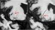
Methods: Patients with acute SSI were prospectively enrolled and categorized into PAD-related SSI and cerebral small-vessel disease (CSVD)-related SSI groups. The imaging features of LSA morphology (branches, length, dilation, and tortuosity), plaques (burden, remodeling index, enhancement degree, and hyperintense plaque), and CSVD (white matter hyperintensity, lacunes, cerebral microbleed, and enlarged perivascular space) were evaluated. The logistic regression was performed to determine the association of dilated LSA with PAD-related SSI and 90-day clinical outcomes.
Results: In total, 131 patients (mean age, 52.2 ± 13.2 years; 99 men) were included. The multivariate logistic regression analysis revealed that the presence of dilated LSAs (odds ratio (OR), 7.40; 95% confidence interval (CI): 1.88-29.17; p = 0.004)) was significantly associated with PAD-related SSI. Moreover, after adjusting for confounding factors, the association of poor outcomes with the total length of LSAs (OR, 0.94; 95% CI: 0.90-0.99; p = 0.011), dilated LSAs (OR, 0.001; 95% CI: 0.0001-0.08; p = 0.002), and plaque burden (OR, 1.35; 95% CI: 1.11-1.63; p = 0.002) remained statistically significant.
Conclusion: The dilation of LSA visualized on WB-VWI could differentiate various subtypes of SSI within LSA territory and was a prognostic imaging marker for 90-day clinical outcomes for PAD-related SSI.
Clinical relevance statement: Evaluation of LSA morphology based on WB-VWI can differentiate the pathogenesis and predict clinical outcomes in SSI, providing crucial insights into the etiologic mechanisms, risk stratification, and tailored therapies for these patients.
Key points: The prognosis of SSIs within lenticulostriate territory depend on the etiology of the disease. LSA dilation on WB-VWI was associated with parental atherosclerosis and better 90-day outcomes. Accurately identifying the etiology of SSIs in lenticulostriate territory assists in treatment decision-making.
期刊介绍:
European Radiology (ER) continuously updates scientific knowledge in radiology by publication of strong original articles and state-of-the-art reviews written by leading radiologists. A well balanced combination of review articles, original papers, short communications from European radiological congresses and information on society matters makes ER an indispensable source for current information in this field.
This is the Journal of the European Society of Radiology, and the official journal of a number of societies.
From 2004-2008 supplements to European Radiology were published under its companion, European Radiology Supplements, ISSN 1613-3749.

 求助内容:
求助内容: 应助结果提醒方式:


