Markus Linder, Lucas Bennink, Richard H. Foxton, Mike Kirkness, Peter D. Westenskow
{"title":"利用胶原杂交肽对小鼠视网膜下活动性纤维化进行体内监测","authors":"Markus Linder, Lucas Bennink, Richard H. Foxton, Mike Kirkness, Peter D. Westenskow","doi":"10.1038/s41684-024-01408-0","DOIUrl":null,"url":null,"abstract":"Subretinal fibrosis is associated with worse visual outcomes in patients with neovascular age-related macular degeneration. As there is a lack of optimal biomarkers and no method that directly detects collagen in the back of the eye, novel tools that monitor fibrosis-related changes in neovascular age-related macular degeneration are needed. Here, using two mouse models (the laser-induced choroidal neovascularization model, and the JR5558 mouse presenting with spontaneous subretinal neovascularization with fibrosis), we imaged active fibrotic lesions using fluorescently labeled collagen hybridizing peptides (CHPs), short peptides that bind to single α-chain collagen structures during collagen remodeling. JR5558 retinal pigment epithelium/choroid flat mounts showed CHP co-staining with fibrosis and epithelial mesenchymal transition-related markers; additionally, CHP histopathology staining correlated with in vivo CHP imaging. After laser-induced choroidal neovascularization, in vivo CHP binding correlated with laser intensity, histopathology CHP and fibronectin staining. Laser-induced choroidal neovascularization showed decreased CHP intensity over time in healing/regressing versus active scars in vivo, whereas increased CHP binding correlated with elevated fibrosis in JR5558 mouse eyes with age. In bispecific angiopoietin 2/vascular endothelial growth factor antibody-treated JR5558 mice, CHPs detected significantly decreased collagen remodeling versus immunoglobulin G control. These results demonstrate the first use of CHPs to directly image remodeling collagen in the eye and as a potential clinical optical biomarker of active subretinal fibrosis associated with ocular neovascularization. This study reports the use of fluorescently labeled collagen hybridizing peptides to directly image collagen remodeling and monitor fibrosis in two mouse models of ocular neovascularization.","PeriodicalId":17936,"journal":{"name":"Lab Animal","volume":null,"pages":null},"PeriodicalIF":5.9000,"publicationDate":"2024-07-26","publicationTypes":"Journal Article","fieldsOfStudy":null,"isOpenAccess":false,"openAccessPdf":"https://www.nature.com/articles/s41684-024-01408-0.pdf","citationCount":"0","resultStr":"{\"title\":\"In vivo monitoring of active subretinal fibrosis in mice using collagen hybridizing peptides\",\"authors\":\"Markus Linder, Lucas Bennink, Richard H. Foxton, Mike Kirkness, Peter D. Westenskow\",\"doi\":\"10.1038/s41684-024-01408-0\",\"DOIUrl\":null,\"url\":null,\"abstract\":\"Subretinal fibrosis is associated with worse visual outcomes in patients with neovascular age-related macular degeneration. As there is a lack of optimal biomarkers and no method that directly detects collagen in the back of the eye, novel tools that monitor fibrosis-related changes in neovascular age-related macular degeneration are needed. Here, using two mouse models (the laser-induced choroidal neovascularization model, and the JR5558 mouse presenting with spontaneous subretinal neovascularization with fibrosis), we imaged active fibrotic lesions using fluorescently labeled collagen hybridizing peptides (CHPs), short peptides that bind to single α-chain collagen structures during collagen remodeling. JR5558 retinal pigment epithelium/choroid flat mounts showed CHP co-staining with fibrosis and epithelial mesenchymal transition-related markers; additionally, CHP histopathology staining correlated with in vivo CHP imaging. After laser-induced choroidal neovascularization, in vivo CHP binding correlated with laser intensity, histopathology CHP and fibronectin staining. Laser-induced choroidal neovascularization showed decreased CHP intensity over time in healing/regressing versus active scars in vivo, whereas increased CHP binding correlated with elevated fibrosis in JR5558 mouse eyes with age. In bispecific angiopoietin 2/vascular endothelial growth factor antibody-treated JR5558 mice, CHPs detected significantly decreased collagen remodeling versus immunoglobulin G control. These results demonstrate the first use of CHPs to directly image remodeling collagen in the eye and as a potential clinical optical biomarker of active subretinal fibrosis associated with ocular neovascularization. This study reports the use of fluorescently labeled collagen hybridizing peptides to directly image collagen remodeling and monitor fibrosis in two mouse models of ocular neovascularization.\",\"PeriodicalId\":17936,\"journal\":{\"name\":\"Lab Animal\",\"volume\":null,\"pages\":null},\"PeriodicalIF\":5.9000,\"publicationDate\":\"2024-07-26\",\"publicationTypes\":\"Journal Article\",\"fieldsOfStudy\":null,\"isOpenAccess\":false,\"openAccessPdf\":\"https://www.nature.com/articles/s41684-024-01408-0.pdf\",\"citationCount\":\"0\",\"resultStr\":null,\"platform\":\"Semanticscholar\",\"paperid\":null,\"PeriodicalName\":\"Lab Animal\",\"FirstCategoryId\":\"97\",\"ListUrlMain\":\"https://www.nature.com/articles/s41684-024-01408-0\",\"RegionNum\":3,\"RegionCategory\":\"农林科学\",\"ArticlePicture\":[],\"TitleCN\":null,\"AbstractTextCN\":null,\"PMCID\":null,\"EPubDate\":\"\",\"PubModel\":\"\",\"JCR\":\"Q1\",\"JCRName\":\"VETERINARY SCIENCES\",\"Score\":null,\"Total\":0}","platform":"Semanticscholar","paperid":null,"PeriodicalName":"Lab Animal","FirstCategoryId":"97","ListUrlMain":"https://www.nature.com/articles/s41684-024-01408-0","RegionNum":3,"RegionCategory":"农林科学","ArticlePicture":[],"TitleCN":null,"AbstractTextCN":null,"PMCID":null,"EPubDate":"","PubModel":"","JCR":"Q1","JCRName":"VETERINARY SCIENCES","Score":null,"Total":0}
In vivo monitoring of active subretinal fibrosis in mice using collagen hybridizing peptides
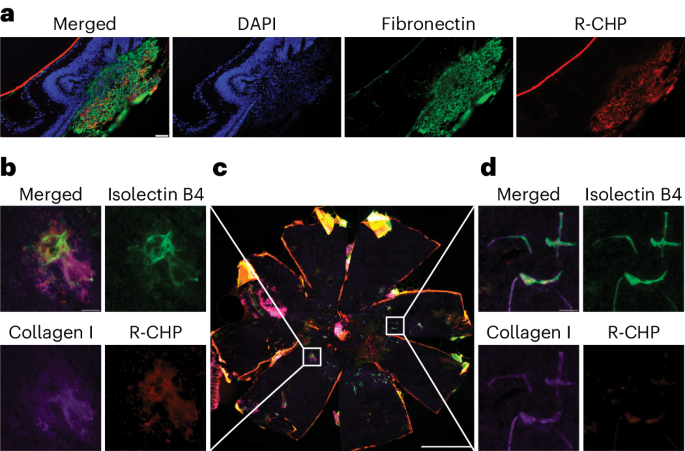
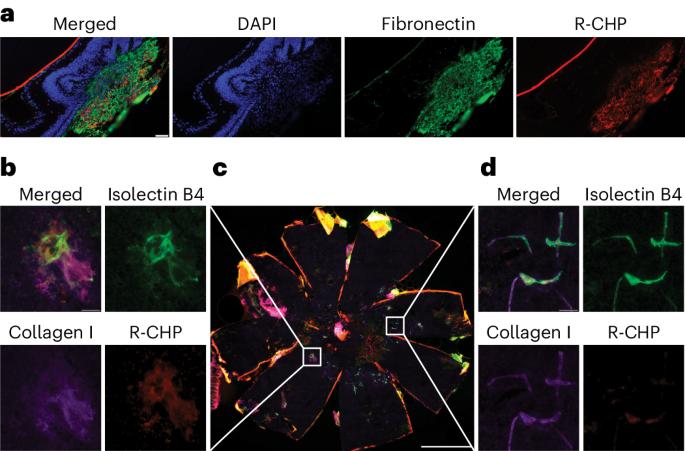
Subretinal fibrosis is associated with worse visual outcomes in patients with neovascular age-related macular degeneration. As there is a lack of optimal biomarkers and no method that directly detects collagen in the back of the eye, novel tools that monitor fibrosis-related changes in neovascular age-related macular degeneration are needed. Here, using two mouse models (the laser-induced choroidal neovascularization model, and the JR5558 mouse presenting with spontaneous subretinal neovascularization with fibrosis), we imaged active fibrotic lesions using fluorescently labeled collagen hybridizing peptides (CHPs), short peptides that bind to single α-chain collagen structures during collagen remodeling. JR5558 retinal pigment epithelium/choroid flat mounts showed CHP co-staining with fibrosis and epithelial mesenchymal transition-related markers; additionally, CHP histopathology staining correlated with in vivo CHP imaging. After laser-induced choroidal neovascularization, in vivo CHP binding correlated with laser intensity, histopathology CHP and fibronectin staining. Laser-induced choroidal neovascularization showed decreased CHP intensity over time in healing/regressing versus active scars in vivo, whereas increased CHP binding correlated with elevated fibrosis in JR5558 mouse eyes with age. In bispecific angiopoietin 2/vascular endothelial growth factor antibody-treated JR5558 mice, CHPs detected significantly decreased collagen remodeling versus immunoglobulin G control. These results demonstrate the first use of CHPs to directly image remodeling collagen in the eye and as a potential clinical optical biomarker of active subretinal fibrosis associated with ocular neovascularization. This study reports the use of fluorescently labeled collagen hybridizing peptides to directly image collagen remodeling and monitor fibrosis in two mouse models of ocular neovascularization.
期刊介绍:
LabAnimal is a Nature Research journal dedicated to in vivo science and technology that improves our basic understanding and use of model organisms of human health and disease. In addition to basic research, methods and technologies, LabAnimal also covers important news, business and regulatory matters that impact the development and application of model organisms for preclinical research.
LabAnimal's focus is on innovative in vivo methods, research and technology covering a wide range of model organisms. Our broad scope ensures that the work we publish reaches the widest possible audience. LabAnimal provides a rigorous and fair peer review of manuscripts, high standards for copyediting and production, and efficient publication.