{"title":"肺外透明肉芽肿:关节内和腱鞘受累的罕见病例。","authors":"Mohanad Alhumayed, Ryan P Austin, Eric Y Chang","doi":"10.1007/s00256-024-04762-9","DOIUrl":null,"url":null,"abstract":"<p><p>Extrapulmonary hyalinizing granuloma (EPHG) is a notably rare condition, representing an exaggerated chronic immune response to antigenic stimuli. This report presents the first documented case of intra-articular and tenosynovial EPHG with radiological evaluation and pathological confirmation in a 60-year-old man presenting with wrist pain and swelling. Imaging findings were relatively symmetric with marked distension of the distal radioulnar joints and extensor tendon sheaths with masses and nodules of various sizes surrounded by synovitis and accompanied by bony erosions. On US, the masses were heterogeneous but mostly hypo- to iso-echoic compared to muscle and relatively hypovascular. On MRI, compared to muscle, the nodules exhibited iso-intense signal on T1-weighted images, iso- to mildly hyper-intense signal on T2-weighted fat-suppressed images, and minimal enhancement on post-contrast images. The diagnosis of EPHG was revealed through biopsy and pathologic examination with glucocorticoids being effective in treatment.</p>","PeriodicalId":21783,"journal":{"name":"Skeletal Radiology","volume":" ","pages":"1119-1124"},"PeriodicalIF":1.9000,"publicationDate":"2025-05-01","publicationTypes":"Journal Article","fieldsOfStudy":null,"isOpenAccess":false,"openAccessPdf":"https://www.ncbi.nlm.nih.gov/pmc/articles/PMC11953202/pdf/","citationCount":"0","resultStr":"{\"title\":\"Extrapulmonary hyalinizing granuloma: a rare case with intra-articular and tenosynovial involvement.\",\"authors\":\"Mohanad Alhumayed, Ryan P Austin, Eric Y Chang\",\"doi\":\"10.1007/s00256-024-04762-9\",\"DOIUrl\":null,\"url\":null,\"abstract\":\"<p><p>Extrapulmonary hyalinizing granuloma (EPHG) is a notably rare condition, representing an exaggerated chronic immune response to antigenic stimuli. This report presents the first documented case of intra-articular and tenosynovial EPHG with radiological evaluation and pathological confirmation in a 60-year-old man presenting with wrist pain and swelling. Imaging findings were relatively symmetric with marked distension of the distal radioulnar joints and extensor tendon sheaths with masses and nodules of various sizes surrounded by synovitis and accompanied by bony erosions. On US, the masses were heterogeneous but mostly hypo- to iso-echoic compared to muscle and relatively hypovascular. On MRI, compared to muscle, the nodules exhibited iso-intense signal on T1-weighted images, iso- to mildly hyper-intense signal on T2-weighted fat-suppressed images, and minimal enhancement on post-contrast images. The diagnosis of EPHG was revealed through biopsy and pathologic examination with glucocorticoids being effective in treatment.</p>\",\"PeriodicalId\":21783,\"journal\":{\"name\":\"Skeletal Radiology\",\"volume\":\" \",\"pages\":\"1119-1124\"},\"PeriodicalIF\":1.9000,\"publicationDate\":\"2025-05-01\",\"publicationTypes\":\"Journal Article\",\"fieldsOfStudy\":null,\"isOpenAccess\":false,\"openAccessPdf\":\"https://www.ncbi.nlm.nih.gov/pmc/articles/PMC11953202/pdf/\",\"citationCount\":\"0\",\"resultStr\":null,\"platform\":\"Semanticscholar\",\"paperid\":null,\"PeriodicalName\":\"Skeletal Radiology\",\"FirstCategoryId\":\"3\",\"ListUrlMain\":\"https://doi.org/10.1007/s00256-024-04762-9\",\"RegionNum\":3,\"RegionCategory\":\"医学\",\"ArticlePicture\":[],\"TitleCN\":null,\"AbstractTextCN\":null,\"PMCID\":null,\"EPubDate\":\"2024/7/24 0:00:00\",\"PubModel\":\"Epub\",\"JCR\":\"Q2\",\"JCRName\":\"ORTHOPEDICS\",\"Score\":null,\"Total\":0}","platform":"Semanticscholar","paperid":null,"PeriodicalName":"Skeletal Radiology","FirstCategoryId":"3","ListUrlMain":"https://doi.org/10.1007/s00256-024-04762-9","RegionNum":3,"RegionCategory":"医学","ArticlePicture":[],"TitleCN":null,"AbstractTextCN":null,"PMCID":null,"EPubDate":"2024/7/24 0:00:00","PubModel":"Epub","JCR":"Q2","JCRName":"ORTHOPEDICS","Score":null,"Total":0}
引用次数: 0
摘要
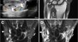
肺外透明肉芽肿(EPHG)是一种非常罕见的疾病,是对抗原刺激的一种夸张的慢性免疫反应。本报告介绍了首例记录在案的关节内和腱鞘外透明肉芽肿病例,该病例是一名 60 岁的男性,因手腕疼痛和肿胀而进行放射学评估和病理学确诊。影像学检查结果相对对称,桡侧远端关节和伸肌腱鞘明显膨胀,有大小不等的肿块和结节,周围有滑膜炎,并伴有骨质侵蚀。在 US 上,肿块呈异质性,但与肌肉相比,大多呈低回声或等回声,血管相对较少。在核磁共振成像上,与肌肉相比,结节在T1加权图像上呈等强信号,在T2加权脂肪抑制图像上呈等强至轻度高强信号,在对比后图像上增强极弱。通过活检和病理检查确诊为 EPHG,糖皮质激素治疗有效。
Extrapulmonary hyalinizing granuloma: a rare case with intra-articular and tenosynovial involvement.
Extrapulmonary hyalinizing granuloma (EPHG) is a notably rare condition, representing an exaggerated chronic immune response to antigenic stimuli. This report presents the first documented case of intra-articular and tenosynovial EPHG with radiological evaluation and pathological confirmation in a 60-year-old man presenting with wrist pain and swelling. Imaging findings were relatively symmetric with marked distension of the distal radioulnar joints and extensor tendon sheaths with masses and nodules of various sizes surrounded by synovitis and accompanied by bony erosions. On US, the masses were heterogeneous but mostly hypo- to iso-echoic compared to muscle and relatively hypovascular. On MRI, compared to muscle, the nodules exhibited iso-intense signal on T1-weighted images, iso- to mildly hyper-intense signal on T2-weighted fat-suppressed images, and minimal enhancement on post-contrast images. The diagnosis of EPHG was revealed through biopsy and pathologic examination with glucocorticoids being effective in treatment.
期刊介绍:
Skeletal Radiology provides a forum for the dissemination of current knowledge and information dealing with disorders of the musculoskeletal system including the spine. While emphasizing the radiological aspects of the many varied skeletal abnormalities, the journal also adopts an interdisciplinary approach, reflecting the membership of the International Skeletal Society. Thus, the anatomical, pathological, physiological, clinical, metabolic and epidemiological aspects of the many entities affecting the skeleton receive appropriate consideration.
This is the Journal of the International Skeletal Society and the Official Journal of the Society of Skeletal Radiology and the Australasian Musculoskelelal Imaging Group.

 求助内容:
求助内容: 应助结果提醒方式:


