A Coudert, L Denis, A Chavignon, S Bodard, M Naveau, P P Sistiaga, R Saulnier, C Orset, D Vivien, C Chappard, O Couture
{"title":"三维经颅超声定位显微镜显示绵羊大脑中的主要动脉。","authors":"A Coudert, L Denis, A Chavignon, S Bodard, M Naveau, P P Sistiaga, R Saulnier, C Orset, D Vivien, C Chappard, O Couture","doi":"10.1109/TUFFC.2024.3432998","DOIUrl":null,"url":null,"abstract":"<p><p>Cerebral circulation ensures the proper functioning of the entire human body, and its interruption, i.e. stroke, leads to irreversible damage. However, tools for observing cerebral circulation are still lacking. Although MRI and CT scans serve as conventional methods, their accessibility remains a challenge, prompting exploration into alternative, portable, and non-ionizing imaging solutions like ultrasound with reduced costs. While Ultrasound Localization Microscopy (ULM) displays potential in high-resolution vessel imaging, its 2D constraints limit its emergency utility. This study delves into the feasibility of 3D ULM with multiplexed probe for transcranial vessel imaging in sheep brains, emulating human skull characteristics. Three sheep underwent 3D ULM imaging, compared with angiographic MRI, while skull characterization was conducted in vivo using ultrashort bone MRI sequences and ex vivo via micro CT. The study showcased 3D ULM's ability to highlight vessels, down to the Circle of Willis, yet within a confined 3D field-of-view. Future enhancements in signal, aberration correction, and human trials hold promise for a portable, volumetric, transcranial ultrasound angiography system.</p>","PeriodicalId":13322,"journal":{"name":"IEEE transactions on ultrasonics, ferroelectrics, and frequency control","volume":"PP ","pages":""},"PeriodicalIF":3.0000,"publicationDate":"2024-07-25","publicationTypes":"Journal Article","fieldsOfStudy":null,"isOpenAccess":false,"openAccessPdf":"","citationCount":"0","resultStr":"{\"title\":\"3D Transcranial ultrasound localization microscopy reveals major arteries in the sheep brain.\",\"authors\":\"A Coudert, L Denis, A Chavignon, S Bodard, M Naveau, P P Sistiaga, R Saulnier, C Orset, D Vivien, C Chappard, O Couture\",\"doi\":\"10.1109/TUFFC.2024.3432998\",\"DOIUrl\":null,\"url\":null,\"abstract\":\"<p><p>Cerebral circulation ensures the proper functioning of the entire human body, and its interruption, i.e. stroke, leads to irreversible damage. However, tools for observing cerebral circulation are still lacking. Although MRI and CT scans serve as conventional methods, their accessibility remains a challenge, prompting exploration into alternative, portable, and non-ionizing imaging solutions like ultrasound with reduced costs. While Ultrasound Localization Microscopy (ULM) displays potential in high-resolution vessel imaging, its 2D constraints limit its emergency utility. This study delves into the feasibility of 3D ULM with multiplexed probe for transcranial vessel imaging in sheep brains, emulating human skull characteristics. Three sheep underwent 3D ULM imaging, compared with angiographic MRI, while skull characterization was conducted in vivo using ultrashort bone MRI sequences and ex vivo via micro CT. The study showcased 3D ULM's ability to highlight vessels, down to the Circle of Willis, yet within a confined 3D field-of-view. Future enhancements in signal, aberration correction, and human trials hold promise for a portable, volumetric, transcranial ultrasound angiography system.</p>\",\"PeriodicalId\":13322,\"journal\":{\"name\":\"IEEE transactions on ultrasonics, ferroelectrics, and frequency control\",\"volume\":\"PP \",\"pages\":\"\"},\"PeriodicalIF\":3.0000,\"publicationDate\":\"2024-07-25\",\"publicationTypes\":\"Journal Article\",\"fieldsOfStudy\":null,\"isOpenAccess\":false,\"openAccessPdf\":\"\",\"citationCount\":\"0\",\"resultStr\":null,\"platform\":\"Semanticscholar\",\"paperid\":null,\"PeriodicalName\":\"IEEE transactions on ultrasonics, ferroelectrics, and frequency control\",\"FirstCategoryId\":\"5\",\"ListUrlMain\":\"https://doi.org/10.1109/TUFFC.2024.3432998\",\"RegionNum\":2,\"RegionCategory\":\"工程技术\",\"ArticlePicture\":[],\"TitleCN\":null,\"AbstractTextCN\":null,\"PMCID\":null,\"EPubDate\":\"\",\"PubModel\":\"\",\"JCR\":\"Q1\",\"JCRName\":\"ACOUSTICS\",\"Score\":null,\"Total\":0}","platform":"Semanticscholar","paperid":null,"PeriodicalName":"IEEE transactions on ultrasonics, ferroelectrics, and frequency control","FirstCategoryId":"5","ListUrlMain":"https://doi.org/10.1109/TUFFC.2024.3432998","RegionNum":2,"RegionCategory":"工程技术","ArticlePicture":[],"TitleCN":null,"AbstractTextCN":null,"PMCID":null,"EPubDate":"","PubModel":"","JCR":"Q1","JCRName":"ACOUSTICS","Score":null,"Total":0}
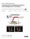
3D Transcranial ultrasound localization microscopy reveals major arteries in the sheep brain.
Cerebral circulation ensures the proper functioning of the entire human body, and its interruption, i.e. stroke, leads to irreversible damage. However, tools for observing cerebral circulation are still lacking. Although MRI and CT scans serve as conventional methods, their accessibility remains a challenge, prompting exploration into alternative, portable, and non-ionizing imaging solutions like ultrasound with reduced costs. While Ultrasound Localization Microscopy (ULM) displays potential in high-resolution vessel imaging, its 2D constraints limit its emergency utility. This study delves into the feasibility of 3D ULM with multiplexed probe for transcranial vessel imaging in sheep brains, emulating human skull characteristics. Three sheep underwent 3D ULM imaging, compared with angiographic MRI, while skull characterization was conducted in vivo using ultrashort bone MRI sequences and ex vivo via micro CT. The study showcased 3D ULM's ability to highlight vessels, down to the Circle of Willis, yet within a confined 3D field-of-view. Future enhancements in signal, aberration correction, and human trials hold promise for a portable, volumetric, transcranial ultrasound angiography system.
期刊介绍:
IEEE Transactions on Ultrasonics, Ferroelectrics and Frequency Control includes the theory, technology, materials, and applications relating to: (1) the generation, transmission, and detection of ultrasonic waves and related phenomena; (2) medical ultrasound, including hyperthermia, bioeffects, tissue characterization and imaging; (3) ferroelectric, piezoelectric, and piezomagnetic materials, including crystals, polycrystalline solids, films, polymers, and composites; (4) frequency control, timing and time distribution, including crystal oscillators and other means of classical frequency control, and atomic, molecular and laser frequency control standards. Areas of interest range from fundamental studies to the design and/or applications of devices and systems.