Farhad Soheilmoghaddam, Hadi Hezaveh, Madeleine Rumble, Justin J Cooper-White
{"title":"在仿骨生物纳米纤维支架中驱动间充质干细胞的骨细胞生成。","authors":"Farhad Soheilmoghaddam, Hadi Hezaveh, Madeleine Rumble, Justin J Cooper-White","doi":"10.1021/acsami.3c14785","DOIUrl":null,"url":null,"abstract":"<p><p>The treatment of critical-sized bone defects caused by tumor removal, skeletal injuries, or infections continues to pose a major clinical challenge. A popular potential alternative solution to autologous bone grafts is a tissue-engineered approach that utilizes the combination of mesenchymal stromal/stem cells (MSCs) with synthetic biomaterial scaffolds. This approach aims to support new bone formation by mimicking many of the biochemical and biophysical cues present within native bone. Regrettably, osteocyte cells, crucial for bone maturation and homeostasis, are rarely produced within MSC-seeded scaffolds, thereby restricting the development of fully mature cortical bone from these synthetic implants. In this work, we have constructed a multimodal scaffold by combining electrospun poly(lactic-<i>co</i>-glycolic acid) (PLGA) fibrous scaffolds with poly(ethylene glycol) (PEG)-based hydrogels that mimic the functional unit of cortical bone, osteon (osteon-mimetic) scaffolds. These scaffolds were decorated with a novel bone morphogenic protein-6 (BMP6) peptide (BMP6p) after our findings revealed that the BMP6p drives higher levels of Smad signaling than the full-length protein counterpart, soluble or when bound to the PEG hydrogel backbone. We show that our osteon-mimetic scaffolds, in presenting concentric layers of BMP6p-PEG hydrogel overlaid on MSC-seeded PLGA nanofibers, promoted the rapid formation of osteocyte-like cells with a phenotypic dendritic morphology, producing early osteocyte markers, including E11/gp38 (E11). Maturation of these osteocyte-like cells was further confirmed by the observation of significant dentin matrix protein 1 (DMP1) throughout our bilayered scaffolds after 3 weeks, even when cultured in a medium without dexamethasone (DEX) or any other osteogenic supplements. These results demonstrate that these osteon-mimetic scaffolds, in presenting biochemical and topographical cues reminiscent of the forming osteon, can drive the formation of osteocyte-like cells <i>in vitro</i> from hBMSCs <i>without</i> the need for any osteogenic factor media supplementation.</p>","PeriodicalId":5,"journal":{"name":"ACS Applied Materials & Interfaces","volume":" ","pages":"40411-40427"},"PeriodicalIF":8.2000,"publicationDate":"2024-08-07","publicationTypes":"Journal Article","fieldsOfStudy":null,"isOpenAccess":false,"openAccessPdf":"","citationCount":"0","resultStr":"{\"title\":\"Driving Osteocytogenesis from Mesenchymal Stem Cells in Osteon-like Biomimetic Nanofibrous Scaffolds.\",\"authors\":\"Farhad Soheilmoghaddam, Hadi Hezaveh, Madeleine Rumble, Justin J Cooper-White\",\"doi\":\"10.1021/acsami.3c14785\",\"DOIUrl\":null,\"url\":null,\"abstract\":\"<p><p>The treatment of critical-sized bone defects caused by tumor removal, skeletal injuries, or infections continues to pose a major clinical challenge. A popular potential alternative solution to autologous bone grafts is a tissue-engineered approach that utilizes the combination of mesenchymal stromal/stem cells (MSCs) with synthetic biomaterial scaffolds. This approach aims to support new bone formation by mimicking many of the biochemical and biophysical cues present within native bone. Regrettably, osteocyte cells, crucial for bone maturation and homeostasis, are rarely produced within MSC-seeded scaffolds, thereby restricting the development of fully mature cortical bone from these synthetic implants. In this work, we have constructed a multimodal scaffold by combining electrospun poly(lactic-<i>co</i>-glycolic acid) (PLGA) fibrous scaffolds with poly(ethylene glycol) (PEG)-based hydrogels that mimic the functional unit of cortical bone, osteon (osteon-mimetic) scaffolds. These scaffolds were decorated with a novel bone morphogenic protein-6 (BMP6) peptide (BMP6p) after our findings revealed that the BMP6p drives higher levels of Smad signaling than the full-length protein counterpart, soluble or when bound to the PEG hydrogel backbone. We show that our osteon-mimetic scaffolds, in presenting concentric layers of BMP6p-PEG hydrogel overlaid on MSC-seeded PLGA nanofibers, promoted the rapid formation of osteocyte-like cells with a phenotypic dendritic morphology, producing early osteocyte markers, including E11/gp38 (E11). Maturation of these osteocyte-like cells was further confirmed by the observation of significant dentin matrix protein 1 (DMP1) throughout our bilayered scaffolds after 3 weeks, even when cultured in a medium without dexamethasone (DEX) or any other osteogenic supplements. These results demonstrate that these osteon-mimetic scaffolds, in presenting biochemical and topographical cues reminiscent of the forming osteon, can drive the formation of osteocyte-like cells <i>in vitro</i> from hBMSCs <i>without</i> the need for any osteogenic factor media supplementation.</p>\",\"PeriodicalId\":5,\"journal\":{\"name\":\"ACS Applied Materials & Interfaces\",\"volume\":\" \",\"pages\":\"40411-40427\"},\"PeriodicalIF\":8.2000,\"publicationDate\":\"2024-08-07\",\"publicationTypes\":\"Journal Article\",\"fieldsOfStudy\":null,\"isOpenAccess\":false,\"openAccessPdf\":\"\",\"citationCount\":\"0\",\"resultStr\":null,\"platform\":\"Semanticscholar\",\"paperid\":null,\"PeriodicalName\":\"ACS Applied Materials & Interfaces\",\"FirstCategoryId\":\"88\",\"ListUrlMain\":\"https://doi.org/10.1021/acsami.3c14785\",\"RegionNum\":2,\"RegionCategory\":\"材料科学\",\"ArticlePicture\":[],\"TitleCN\":null,\"AbstractTextCN\":null,\"PMCID\":null,\"EPubDate\":\"2024/7/23 0:00:00\",\"PubModel\":\"Epub\",\"JCR\":\"Q1\",\"JCRName\":\"MATERIALS SCIENCE, MULTIDISCIPLINARY\",\"Score\":null,\"Total\":0}","platform":"Semanticscholar","paperid":null,"PeriodicalName":"ACS Applied Materials & Interfaces","FirstCategoryId":"88","ListUrlMain":"https://doi.org/10.1021/acsami.3c14785","RegionNum":2,"RegionCategory":"材料科学","ArticlePicture":[],"TitleCN":null,"AbstractTextCN":null,"PMCID":null,"EPubDate":"2024/7/23 0:00:00","PubModel":"Epub","JCR":"Q1","JCRName":"MATERIALS SCIENCE, MULTIDISCIPLINARY","Score":null,"Total":0}
Driving Osteocytogenesis from Mesenchymal Stem Cells in Osteon-like Biomimetic Nanofibrous Scaffolds.
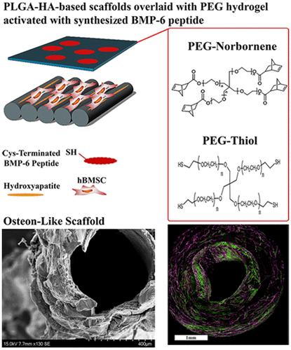
The treatment of critical-sized bone defects caused by tumor removal, skeletal injuries, or infections continues to pose a major clinical challenge. A popular potential alternative solution to autologous bone grafts is a tissue-engineered approach that utilizes the combination of mesenchymal stromal/stem cells (MSCs) with synthetic biomaterial scaffolds. This approach aims to support new bone formation by mimicking many of the biochemical and biophysical cues present within native bone. Regrettably, osteocyte cells, crucial for bone maturation and homeostasis, are rarely produced within MSC-seeded scaffolds, thereby restricting the development of fully mature cortical bone from these synthetic implants. In this work, we have constructed a multimodal scaffold by combining electrospun poly(lactic-co-glycolic acid) (PLGA) fibrous scaffolds with poly(ethylene glycol) (PEG)-based hydrogels that mimic the functional unit of cortical bone, osteon (osteon-mimetic) scaffolds. These scaffolds were decorated with a novel bone morphogenic protein-6 (BMP6) peptide (BMP6p) after our findings revealed that the BMP6p drives higher levels of Smad signaling than the full-length protein counterpart, soluble or when bound to the PEG hydrogel backbone. We show that our osteon-mimetic scaffolds, in presenting concentric layers of BMP6p-PEG hydrogel overlaid on MSC-seeded PLGA nanofibers, promoted the rapid formation of osteocyte-like cells with a phenotypic dendritic morphology, producing early osteocyte markers, including E11/gp38 (E11). Maturation of these osteocyte-like cells was further confirmed by the observation of significant dentin matrix protein 1 (DMP1) throughout our bilayered scaffolds after 3 weeks, even when cultured in a medium without dexamethasone (DEX) or any other osteogenic supplements. These results demonstrate that these osteon-mimetic scaffolds, in presenting biochemical and topographical cues reminiscent of the forming osteon, can drive the formation of osteocyte-like cells in vitro from hBMSCs without the need for any osteogenic factor media supplementation.
期刊介绍:
ACS Applied Materials & Interfaces is a leading interdisciplinary journal that brings together chemists, engineers, physicists, and biologists to explore the development and utilization of newly-discovered materials and interfacial processes for specific applications. Our journal has experienced remarkable growth since its establishment in 2009, both in terms of the number of articles published and the impact of the research showcased. We are proud to foster a truly global community, with the majority of published articles originating from outside the United States, reflecting the rapid growth of applied research worldwide.
 求助内容:
求助内容: 应助结果提醒方式:


