Sami F Alaraj, Samuel O Krider, Ahmed Elsayes, Eseosa Bazuaye, Glenn M Garcia
{"title":"双侧前臂嗜血性假瘤:一份独特的病例报告。","authors":"Sami F Alaraj, Samuel O Krider, Ahmed Elsayes, Eseosa Bazuaye, Glenn M Garcia","doi":"10.1007/s00256-024-04760-x","DOIUrl":null,"url":null,"abstract":"<p><p>Hemophilic pseudotumor (HP) is a rarely encountered cystic mass that forms as a result of repeated bleeding from extra-articular soft tissues. HP cases have been previously documented in several locations in the body, most commonly in the femur and pelvis. To date, no upper extremity case involving the bilateral forearms has been reported. The current case involves an adult male with uncontrolled hemophilia who presented with diffuse enlargement of the bilateral forearms with associated pain. Radiographs and magnetic resonance imaging (MRI) were subsequently performed revealing variable aged hemorrhagic, expansile, lytic intramedullary lesions. In keeping with the history, a subsequent radiologic diagnosis of HP was favored, among other differentials, including benign and malignant processes with biopsy confirming the diagnosis. The hemorrhagic masses were surgically excised after initial management with factor VIII replacement. This case details a unique presentation of this pathology in the bilateral forearms and highlights the diagnostic value of radiographs and MRI in diagnosis and management.</p>","PeriodicalId":21783,"journal":{"name":"Skeletal Radiology","volume":" ","pages":"1109-1117"},"PeriodicalIF":2.2000,"publicationDate":"2025-05-01","publicationTypes":"Journal Article","fieldsOfStudy":null,"isOpenAccess":false,"openAccessPdf":"","citationCount":"0","resultStr":"{\"title\":\"Hemophilic pseudotumor in the bilateral forearms: a unique case report.\",\"authors\":\"Sami F Alaraj, Samuel O Krider, Ahmed Elsayes, Eseosa Bazuaye, Glenn M Garcia\",\"doi\":\"10.1007/s00256-024-04760-x\",\"DOIUrl\":null,\"url\":null,\"abstract\":\"<p><p>Hemophilic pseudotumor (HP) is a rarely encountered cystic mass that forms as a result of repeated bleeding from extra-articular soft tissues. HP cases have been previously documented in several locations in the body, most commonly in the femur and pelvis. To date, no upper extremity case involving the bilateral forearms has been reported. The current case involves an adult male with uncontrolled hemophilia who presented with diffuse enlargement of the bilateral forearms with associated pain. Radiographs and magnetic resonance imaging (MRI) were subsequently performed revealing variable aged hemorrhagic, expansile, lytic intramedullary lesions. In keeping with the history, a subsequent radiologic diagnosis of HP was favored, among other differentials, including benign and malignant processes with biopsy confirming the diagnosis. The hemorrhagic masses were surgically excised after initial management with factor VIII replacement. This case details a unique presentation of this pathology in the bilateral forearms and highlights the diagnostic value of radiographs and MRI in diagnosis and management.</p>\",\"PeriodicalId\":21783,\"journal\":{\"name\":\"Skeletal Radiology\",\"volume\":\" \",\"pages\":\"1109-1117\"},\"PeriodicalIF\":2.2000,\"publicationDate\":\"2025-05-01\",\"publicationTypes\":\"Journal Article\",\"fieldsOfStudy\":null,\"isOpenAccess\":false,\"openAccessPdf\":\"\",\"citationCount\":\"0\",\"resultStr\":null,\"platform\":\"Semanticscholar\",\"paperid\":null,\"PeriodicalName\":\"Skeletal Radiology\",\"FirstCategoryId\":\"3\",\"ListUrlMain\":\"https://doi.org/10.1007/s00256-024-04760-x\",\"RegionNum\":3,\"RegionCategory\":\"医学\",\"ArticlePicture\":[],\"TitleCN\":null,\"AbstractTextCN\":null,\"PMCID\":null,\"EPubDate\":\"2024/7/23 0:00:00\",\"PubModel\":\"Epub\",\"JCR\":\"Q2\",\"JCRName\":\"ORTHOPEDICS\",\"Score\":null,\"Total\":0}","platform":"Semanticscholar","paperid":null,"PeriodicalName":"Skeletal Radiology","FirstCategoryId":"3","ListUrlMain":"https://doi.org/10.1007/s00256-024-04760-x","RegionNum":3,"RegionCategory":"医学","ArticlePicture":[],"TitleCN":null,"AbstractTextCN":null,"PMCID":null,"EPubDate":"2024/7/23 0:00:00","PubModel":"Epub","JCR":"Q2","JCRName":"ORTHOPEDICS","Score":null,"Total":0}
Hemophilic pseudotumor in the bilateral forearms: a unique case report.
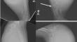
Hemophilic pseudotumor (HP) is a rarely encountered cystic mass that forms as a result of repeated bleeding from extra-articular soft tissues. HP cases have been previously documented in several locations in the body, most commonly in the femur and pelvis. To date, no upper extremity case involving the bilateral forearms has been reported. The current case involves an adult male with uncontrolled hemophilia who presented with diffuse enlargement of the bilateral forearms with associated pain. Radiographs and magnetic resonance imaging (MRI) were subsequently performed revealing variable aged hemorrhagic, expansile, lytic intramedullary lesions. In keeping with the history, a subsequent radiologic diagnosis of HP was favored, among other differentials, including benign and malignant processes with biopsy confirming the diagnosis. The hemorrhagic masses were surgically excised after initial management with factor VIII replacement. This case details a unique presentation of this pathology in the bilateral forearms and highlights the diagnostic value of radiographs and MRI in diagnosis and management.
期刊介绍:
Skeletal Radiology provides a forum for the dissemination of current knowledge and information dealing with disorders of the musculoskeletal system including the spine. While emphasizing the radiological aspects of the many varied skeletal abnormalities, the journal also adopts an interdisciplinary approach, reflecting the membership of the International Skeletal Society. Thus, the anatomical, pathological, physiological, clinical, metabolic and epidemiological aspects of the many entities affecting the skeleton receive appropriate consideration.
This is the Journal of the International Skeletal Society and the Official Journal of the Society of Skeletal Radiology and the Australasian Musculoskelelal Imaging Group.

 求助内容:
求助内容: 应助结果提醒方式:


