Marcelo Bordalo, Patricia Nunez de Aysa, Paulo Victor Partezani Helito, Mohamed Abdelatif Djadoun, Maria Lua Sampaio Gulde, Juan Manuel Alonso
{"title":"青少年足球运动员半膜肌和内收肌的肌肉内伤。","authors":"Marcelo Bordalo, Patricia Nunez de Aysa, Paulo Victor Partezani Helito, Mohamed Abdelatif Djadoun, Maria Lua Sampaio Gulde, Juan Manuel Alonso","doi":"10.1007/s00256-024-04757-6","DOIUrl":null,"url":null,"abstract":"<p><p>Degloving muscle injury was described for the rectus femoris where the inner bipennate component is dissociated from its superficial unipennate component. The semimembranosus muscle displays a distinctive dual morphology, featuring both unipennate and bipennate muscle fibers. Nevertheless, this specific tear pattern has not been previously documented. Conversely, the adductor longus muscle showcases an elongated intramuscular tendon segment, indicating a multipennate morphology. We present two separate cases of previous undescribed degloving injuries of the semimembranosus and the adductor longus in teenage soccer players with MRI and ultrasound diagnosis, ultrasound-guided hematoma aspiration, and recovery timelines for return-to-play.</p>","PeriodicalId":21783,"journal":{"name":"Skeletal Radiology","volume":" ","pages":"887-892"},"PeriodicalIF":1.9000,"publicationDate":"2025-04-01","publicationTypes":"Journal Article","fieldsOfStudy":null,"isOpenAccess":false,"openAccessPdf":"https://www.ncbi.nlm.nih.gov/pmc/articles/PMC11845424/pdf/","citationCount":"0","resultStr":"{\"title\":\"Degloving intramuscular injuries of the semimembranosus and adductor longus muscles in adolescent soccer players.\",\"authors\":\"Marcelo Bordalo, Patricia Nunez de Aysa, Paulo Victor Partezani Helito, Mohamed Abdelatif Djadoun, Maria Lua Sampaio Gulde, Juan Manuel Alonso\",\"doi\":\"10.1007/s00256-024-04757-6\",\"DOIUrl\":null,\"url\":null,\"abstract\":\"<p><p>Degloving muscle injury was described for the rectus femoris where the inner bipennate component is dissociated from its superficial unipennate component. The semimembranosus muscle displays a distinctive dual morphology, featuring both unipennate and bipennate muscle fibers. Nevertheless, this specific tear pattern has not been previously documented. Conversely, the adductor longus muscle showcases an elongated intramuscular tendon segment, indicating a multipennate morphology. We present two separate cases of previous undescribed degloving injuries of the semimembranosus and the adductor longus in teenage soccer players with MRI and ultrasound diagnosis, ultrasound-guided hematoma aspiration, and recovery timelines for return-to-play.</p>\",\"PeriodicalId\":21783,\"journal\":{\"name\":\"Skeletal Radiology\",\"volume\":\" \",\"pages\":\"887-892\"},\"PeriodicalIF\":1.9000,\"publicationDate\":\"2025-04-01\",\"publicationTypes\":\"Journal Article\",\"fieldsOfStudy\":null,\"isOpenAccess\":false,\"openAccessPdf\":\"https://www.ncbi.nlm.nih.gov/pmc/articles/PMC11845424/pdf/\",\"citationCount\":\"0\",\"resultStr\":null,\"platform\":\"Semanticscholar\",\"paperid\":null,\"PeriodicalName\":\"Skeletal Radiology\",\"FirstCategoryId\":\"3\",\"ListUrlMain\":\"https://doi.org/10.1007/s00256-024-04757-6\",\"RegionNum\":3,\"RegionCategory\":\"医学\",\"ArticlePicture\":[],\"TitleCN\":null,\"AbstractTextCN\":null,\"PMCID\":null,\"EPubDate\":\"2024/7/23 0:00:00\",\"PubModel\":\"Epub\",\"JCR\":\"Q2\",\"JCRName\":\"ORTHOPEDICS\",\"Score\":null,\"Total\":0}","platform":"Semanticscholar","paperid":null,"PeriodicalName":"Skeletal Radiology","FirstCategoryId":"3","ListUrlMain":"https://doi.org/10.1007/s00256-024-04757-6","RegionNum":3,"RegionCategory":"医学","ArticlePicture":[],"TitleCN":null,"AbstractTextCN":null,"PMCID":null,"EPubDate":"2024/7/23 0:00:00","PubModel":"Epub","JCR":"Q2","JCRName":"ORTHOPEDICS","Score":null,"Total":0}
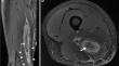
Degloving intramuscular injuries of the semimembranosus and adductor longus muscles in adolescent soccer players.
Degloving muscle injury was described for the rectus femoris where the inner bipennate component is dissociated from its superficial unipennate component. The semimembranosus muscle displays a distinctive dual morphology, featuring both unipennate and bipennate muscle fibers. Nevertheless, this specific tear pattern has not been previously documented. Conversely, the adductor longus muscle showcases an elongated intramuscular tendon segment, indicating a multipennate morphology. We present two separate cases of previous undescribed degloving injuries of the semimembranosus and the adductor longus in teenage soccer players with MRI and ultrasound diagnosis, ultrasound-guided hematoma aspiration, and recovery timelines for return-to-play.
期刊介绍:
Skeletal Radiology provides a forum for the dissemination of current knowledge and information dealing with disorders of the musculoskeletal system including the spine. While emphasizing the radiological aspects of the many varied skeletal abnormalities, the journal also adopts an interdisciplinary approach, reflecting the membership of the International Skeletal Society. Thus, the anatomical, pathological, physiological, clinical, metabolic and epidemiological aspects of the many entities affecting the skeleton receive appropriate consideration.
This is the Journal of the International Skeletal Society and the Official Journal of the Society of Skeletal Radiology and the Australasian Musculoskelelal Imaging Group.

 求助内容:
求助内容: 应助结果提醒方式:


