{"title":"用于区分病理确诊的皮质基底层变性及其模拟病例的常规磁共振成像关键特征:J-VAC 研究的回顾性分析。","authors":"Keita Sakurai, Aya M Tokumaru, Mari Yoshida, Yuko Saito, Koichi Wakabayashi, Takashi Komori, Masato Hasegawa, Takeshi Ikeuchi, Yuichi Hayashi, Takayoshi Shimohata, Shigeo Murayama, Yasushi Iwasaki, Toshiki Uchihara, Motoko Sakai, Ichiro Yabe, Satoshi Tanikawa, Hiroshi Takigawa, Tadashi Adachi, Ritsuko Hanajima, Harutoshi Fujimura, Kentaro Hayashi, Keizo Sugaya, Kazuko Hasegawa, Terunori Sano, Masaki Takao, Osamu Yokota, Tomoko Miki, Michio Kobayashi, Nobutaka Arai, Takuya Ohkubo, Takanori Yokota, Keiko Mori, Masumi Ito, Chiho Ishida, Jiro Idezuka, Yasuko Toyoshima, Masato Kanazawa, Masashi Aoki, Takafumi Hasegawa, Hirohisa Watanabe, Atsushi Hashizume, Hisayoshi Niwa, Keizo Yasui, Keita Ito, Yukihiko Washimi, Akatsuki Kubota, Tatsushi Toda, Kenji Nakashima, Ikuko Aiba","doi":"10.1007/s00234-024-03432-w","DOIUrl":null,"url":null,"abstract":"<p><strong>Purpose: </strong>Due to the indistinguishable clinical features of corticobasal syndrome (CBS), the antemortem differentiation between corticobasal degeneration (CBD) and its mimics remains challenging. However, the utility of conventional magnetic resonance imaging (MRI) for the diagnosis of CBD has not been sufficiently evaluated. This study aimed to investigate the diagnostic performance of conventional MRI findings in differentiating pathologically confirmed CBD from its mimics.</p><p><strong>Methods: </strong>Semiquantitative visual rating scales were employed to assess the degree and distribution of atrophy and asymmetry on conventional T1-weighted and T2-weighted images. Additionally, subcortical white matter hyperintensity (SWMH) on fluid-attenuated inversion recovery images were visually evaluated.</p><p><strong>Results: </strong>In addition to 19 patients with CBD, 16 with CBD mimics (progressive supranuclear palsy (PSP): 9, Alzheimer's disease (AD): 4, dementia with Lewy bodies (DLB): 1, frontotemporal lobar degeneration with TAR DNA-binding protein of 43 kDa(FTLD-TDP): 1, and globular glial tauopathy (GGT): 1) were investigated. Compared with the CBD group, the PSP-CBS subgroup showed severe midbrain atrophy without SWMH. The non-PSP-CBS subgroup, comprising patients with AD, DLB, FTLD-TDP, and GGT, showed severe temporal atrophy with widespread asymmetry, especially in the temporal lobes. In addition to over half of the patients with CBD, two with FTLD-TDP and GGT showed SWMH, respectively.</p><p><strong>Conclusion: </strong>This study elucidates the distinct structural changes between the CBD and its mimics based on visual rating scales. The evaluation of atrophic distribution and SWMH may serve as imaging biomarkers of conventional MRI for detecting background pathologies.</p>","PeriodicalId":19422,"journal":{"name":"Neuroradiology","volume":" ","pages":"1917-1929"},"PeriodicalIF":2.4000,"publicationDate":"2024-11-01","publicationTypes":"Journal Article","fieldsOfStudy":null,"isOpenAccess":false,"openAccessPdf":"https://www.ncbi.nlm.nih.gov/pmc/articles/PMC11535003/pdf/","citationCount":"0","resultStr":"{\"title\":\"Conventional magnetic resonance imaging key features for distinguishing pathologically confirmed corticobasal degeneration from its mimics: a retrospective analysis of the J-VAC study.\",\"authors\":\"Keita Sakurai, Aya M Tokumaru, Mari Yoshida, Yuko Saito, Koichi Wakabayashi, Takashi Komori, Masato Hasegawa, Takeshi Ikeuchi, Yuichi Hayashi, Takayoshi Shimohata, Shigeo Murayama, Yasushi Iwasaki, Toshiki Uchihara, Motoko Sakai, Ichiro Yabe, Satoshi Tanikawa, Hiroshi Takigawa, Tadashi Adachi, Ritsuko Hanajima, Harutoshi Fujimura, Kentaro Hayashi, Keizo Sugaya, Kazuko Hasegawa, Terunori Sano, Masaki Takao, Osamu Yokota, Tomoko Miki, Michio Kobayashi, Nobutaka Arai, Takuya Ohkubo, Takanori Yokota, Keiko Mori, Masumi Ito, Chiho Ishida, Jiro Idezuka, Yasuko Toyoshima, Masato Kanazawa, Masashi Aoki, Takafumi Hasegawa, Hirohisa Watanabe, Atsushi Hashizume, Hisayoshi Niwa, Keizo Yasui, Keita Ito, Yukihiko Washimi, Akatsuki Kubota, Tatsushi Toda, Kenji Nakashima, Ikuko Aiba\",\"doi\":\"10.1007/s00234-024-03432-w\",\"DOIUrl\":null,\"url\":null,\"abstract\":\"<p><strong>Purpose: </strong>Due to the indistinguishable clinical features of corticobasal syndrome (CBS), the antemortem differentiation between corticobasal degeneration (CBD) and its mimics remains challenging. However, the utility of conventional magnetic resonance imaging (MRI) for the diagnosis of CBD has not been sufficiently evaluated. This study aimed to investigate the diagnostic performance of conventional MRI findings in differentiating pathologically confirmed CBD from its mimics.</p><p><strong>Methods: </strong>Semiquantitative visual rating scales were employed to assess the degree and distribution of atrophy and asymmetry on conventional T1-weighted and T2-weighted images. Additionally, subcortical white matter hyperintensity (SWMH) on fluid-attenuated inversion recovery images were visually evaluated.</p><p><strong>Results: </strong>In addition to 19 patients with CBD, 16 with CBD mimics (progressive supranuclear palsy (PSP): 9, Alzheimer's disease (AD): 4, dementia with Lewy bodies (DLB): 1, frontotemporal lobar degeneration with TAR DNA-binding protein of 43 kDa(FTLD-TDP): 1, and globular glial tauopathy (GGT): 1) were investigated. Compared with the CBD group, the PSP-CBS subgroup showed severe midbrain atrophy without SWMH. The non-PSP-CBS subgroup, comprising patients with AD, DLB, FTLD-TDP, and GGT, showed severe temporal atrophy with widespread asymmetry, especially in the temporal lobes. In addition to over half of the patients with CBD, two with FTLD-TDP and GGT showed SWMH, respectively.</p><p><strong>Conclusion: </strong>This study elucidates the distinct structural changes between the CBD and its mimics based on visual rating scales. The evaluation of atrophic distribution and SWMH may serve as imaging biomarkers of conventional MRI for detecting background pathologies.</p>\",\"PeriodicalId\":19422,\"journal\":{\"name\":\"Neuroradiology\",\"volume\":\" \",\"pages\":\"1917-1929\"},\"PeriodicalIF\":2.4000,\"publicationDate\":\"2024-11-01\",\"publicationTypes\":\"Journal Article\",\"fieldsOfStudy\":null,\"isOpenAccess\":false,\"openAccessPdf\":\"https://www.ncbi.nlm.nih.gov/pmc/articles/PMC11535003/pdf/\",\"citationCount\":\"0\",\"resultStr\":null,\"platform\":\"Semanticscholar\",\"paperid\":null,\"PeriodicalName\":\"Neuroradiology\",\"FirstCategoryId\":\"3\",\"ListUrlMain\":\"https://doi.org/10.1007/s00234-024-03432-w\",\"RegionNum\":3,\"RegionCategory\":\"医学\",\"ArticlePicture\":[],\"TitleCN\":null,\"AbstractTextCN\":null,\"PMCID\":null,\"EPubDate\":\"2024/7/22 0:00:00\",\"PubModel\":\"Epub\",\"JCR\":\"Q2\",\"JCRName\":\"CLINICAL NEUROLOGY\",\"Score\":null,\"Total\":0}","platform":"Semanticscholar","paperid":null,"PeriodicalName":"Neuroradiology","FirstCategoryId":"3","ListUrlMain":"https://doi.org/10.1007/s00234-024-03432-w","RegionNum":3,"RegionCategory":"医学","ArticlePicture":[],"TitleCN":null,"AbstractTextCN":null,"PMCID":null,"EPubDate":"2024/7/22 0:00:00","PubModel":"Epub","JCR":"Q2","JCRName":"CLINICAL NEUROLOGY","Score":null,"Total":0}
Conventional magnetic resonance imaging key features for distinguishing pathologically confirmed corticobasal degeneration from its mimics: a retrospective analysis of the J-VAC study.
Purpose: Due to the indistinguishable clinical features of corticobasal syndrome (CBS), the antemortem differentiation between corticobasal degeneration (CBD) and its mimics remains challenging. However, the utility of conventional magnetic resonance imaging (MRI) for the diagnosis of CBD has not been sufficiently evaluated. This study aimed to investigate the diagnostic performance of conventional MRI findings in differentiating pathologically confirmed CBD from its mimics.
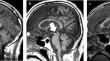
Methods: Semiquantitative visual rating scales were employed to assess the degree and distribution of atrophy and asymmetry on conventional T1-weighted and T2-weighted images. Additionally, subcortical white matter hyperintensity (SWMH) on fluid-attenuated inversion recovery images were visually evaluated.
Results: In addition to 19 patients with CBD, 16 with CBD mimics (progressive supranuclear palsy (PSP): 9, Alzheimer's disease (AD): 4, dementia with Lewy bodies (DLB): 1, frontotemporal lobar degeneration with TAR DNA-binding protein of 43 kDa(FTLD-TDP): 1, and globular glial tauopathy (GGT): 1) were investigated. Compared with the CBD group, the PSP-CBS subgroup showed severe midbrain atrophy without SWMH. The non-PSP-CBS subgroup, comprising patients with AD, DLB, FTLD-TDP, and GGT, showed severe temporal atrophy with widespread asymmetry, especially in the temporal lobes. In addition to over half of the patients with CBD, two with FTLD-TDP and GGT showed SWMH, respectively.
Conclusion: This study elucidates the distinct structural changes between the CBD and its mimics based on visual rating scales. The evaluation of atrophic distribution and SWMH may serve as imaging biomarkers of conventional MRI for detecting background pathologies.
期刊介绍:
Neuroradiology aims to provide state-of-the-art medical and scientific information in the fields of Neuroradiology, Neurosciences, Neurology, Psychiatry, Neurosurgery, and related medical specialities. Neuroradiology as the official Journal of the European Society of Neuroradiology receives submissions from all parts of the world and publishes peer-reviewed original research, comprehensive reviews, educational papers, opinion papers, and short reports on exceptional clinical observations and new technical developments in the field of Neuroimaging and Neurointervention. The journal has subsections for Diagnostic and Interventional Neuroradiology, Advanced Neuroimaging, Paediatric Neuroradiology, Head-Neck-ENT Radiology, Spine Neuroradiology, and for submissions from Japan. Neuroradiology aims to provide new knowledge about and insights into the function and pathology of the human nervous system that may help to better diagnose and treat nervous system diseases. Neuroradiology is a member of the Committee on Publication Ethics (COPE) and follows the COPE core practices. Neuroradiology prefers articles that are free of bias, self-critical regarding limitations, transparent and clear in describing study participants, methods, and statistics, and short in presenting results. Before peer-review all submissions are automatically checked by iThenticate to assess for potential overlap in prior publication.

 求助内容:
求助内容: 应助结果提醒方式:


