Zhi-Hua Li, Xing-Chi Liu, Dan Wang, Zhi-Ling Zhang, Gang Chen, Zi-Li Yu and Zhi-Quan Tian
{"title":"使用超亮荧光纳米球试纸同时检测两种亚型细胞外囊泡。","authors":"Zhi-Hua Li, Xing-Chi Liu, Dan Wang, Zhi-Ling Zhang, Gang Chen, Zi-Li Yu and Zhi-Quan Tian","doi":"10.1039/D4AY00712C","DOIUrl":null,"url":null,"abstract":"<p >In recent years, the cargo profiles of extracellular vesicles (EVs), which were inherited from their parent cells, have emerged as a reliable biomarker for liquid biopsy (LB) in disease diagnosis, prognosis, and treatment monitoring. EVs secreted by different cells exhibit distinct characteristics, particularly in terms of disease diagnosis and prediction. However, currently available techniques for the quantitative analysis of EV cargoes, including enzyme-linked immunosorbent assay (ELISA), cannot specifically identify the cellular origin of EVs, thus seriously affecting the accuracy of EV-based liquid biopsy. In light of this, we here developed ultrabright fluorescent nanosphere (FNs)-based test strips which have the unique capability to specifically assess the levels of PD-L1-positive EVs (PD-L1<small><sup>+</sup></small> EVs) derived from both tumor cells and immune cells in bodily fluids. The levels of PD-L1<small><sup>+</sup></small> EV subpopulations in human saliva were quantified using the ultrabright fluorescent nanosphere-based test strips with more convenience and higher efficiency (detection time <30 min). Results demonstrated that the fluorescence intensity of the test line exhibited a good linear relationship respectively with the PD-L1 levels of tumor cell- (<em>R</em><small><sup>2</sup></small> = 0.993) and immune cell-derived EVs (<em>R</em><small><sup>2</sup></small> = 0.982) in human saliva. By assessing the levels of PD-L1<small><sup>+</sup></small> EV subpopulations, our test strips hold immense potential for advancing the application of PD-L1<small><sup>+</sup></small> EV subpopulation-based predictions in tumor diagnosis and prognosis evaluation. In summary, by integrating the benefits of FNs and lateral flow chromatography, we here provide a strategy to accurately measure the cargo levels of EVs originating from diverse cell sources in bodily fluids.</p>","PeriodicalId":64,"journal":{"name":"Analytical Methods","volume":" 31","pages":" 5403-5411"},"PeriodicalIF":2.6000,"publicationDate":"2024-07-22","publicationTypes":"Journal Article","fieldsOfStudy":null,"isOpenAccess":false,"openAccessPdf":"","citationCount":"0","resultStr":"{\"title\":\"Simultaneous detection of two subtypes of extracellular vesicles using ultrabright fluorescent nanosphere-based test strips†\",\"authors\":\"Zhi-Hua Li, Xing-Chi Liu, Dan Wang, Zhi-Ling Zhang, Gang Chen, Zi-Li Yu and Zhi-Quan Tian\",\"doi\":\"10.1039/D4AY00712C\",\"DOIUrl\":null,\"url\":null,\"abstract\":\"<p >In recent years, the cargo profiles of extracellular vesicles (EVs), which were inherited from their parent cells, have emerged as a reliable biomarker for liquid biopsy (LB) in disease diagnosis, prognosis, and treatment monitoring. EVs secreted by different cells exhibit distinct characteristics, particularly in terms of disease diagnosis and prediction. However, currently available techniques for the quantitative analysis of EV cargoes, including enzyme-linked immunosorbent assay (ELISA), cannot specifically identify the cellular origin of EVs, thus seriously affecting the accuracy of EV-based liquid biopsy. In light of this, we here developed ultrabright fluorescent nanosphere (FNs)-based test strips which have the unique capability to specifically assess the levels of PD-L1-positive EVs (PD-L1<small><sup>+</sup></small> EVs) derived from both tumor cells and immune cells in bodily fluids. The levels of PD-L1<small><sup>+</sup></small> EV subpopulations in human saliva were quantified using the ultrabright fluorescent nanosphere-based test strips with more convenience and higher efficiency (detection time <30 min). Results demonstrated that the fluorescence intensity of the test line exhibited a good linear relationship respectively with the PD-L1 levels of tumor cell- (<em>R</em><small><sup>2</sup></small> = 0.993) and immune cell-derived EVs (<em>R</em><small><sup>2</sup></small> = 0.982) in human saliva. By assessing the levels of PD-L1<small><sup>+</sup></small> EV subpopulations, our test strips hold immense potential for advancing the application of PD-L1<small><sup>+</sup></small> EV subpopulation-based predictions in tumor diagnosis and prognosis evaluation. In summary, by integrating the benefits of FNs and lateral flow chromatography, we here provide a strategy to accurately measure the cargo levels of EVs originating from diverse cell sources in bodily fluids.</p>\",\"PeriodicalId\":64,\"journal\":{\"name\":\"Analytical Methods\",\"volume\":\" 31\",\"pages\":\" 5403-5411\"},\"PeriodicalIF\":2.6000,\"publicationDate\":\"2024-07-22\",\"publicationTypes\":\"Journal Article\",\"fieldsOfStudy\":null,\"isOpenAccess\":false,\"openAccessPdf\":\"\",\"citationCount\":\"0\",\"resultStr\":null,\"platform\":\"Semanticscholar\",\"paperid\":null,\"PeriodicalName\":\"Analytical Methods\",\"FirstCategoryId\":\"92\",\"ListUrlMain\":\"https://pubs.rsc.org/en/content/articlelanding/2024/ay/d4ay00712c\",\"RegionNum\":3,\"RegionCategory\":\"化学\",\"ArticlePicture\":[],\"TitleCN\":null,\"AbstractTextCN\":null,\"PMCID\":null,\"EPubDate\":\"\",\"PubModel\":\"\",\"JCR\":\"Q2\",\"JCRName\":\"CHEMISTRY, ANALYTICAL\",\"Score\":null,\"Total\":0}","platform":"Semanticscholar","paperid":null,"PeriodicalName":"Analytical Methods","FirstCategoryId":"92","ListUrlMain":"https://pubs.rsc.org/en/content/articlelanding/2024/ay/d4ay00712c","RegionNum":3,"RegionCategory":"化学","ArticlePicture":[],"TitleCN":null,"AbstractTextCN":null,"PMCID":null,"EPubDate":"","PubModel":"","JCR":"Q2","JCRName":"CHEMISTRY, ANALYTICAL","Score":null,"Total":0}
引用次数: 0
摘要
近年来,细胞外囊泡(EVs)从其母细胞继承下来的货物特征已成为液体活检(LB)在疾病诊断、预后和治疗监测方面的可靠生物标志物。不同细胞分泌的 EVs 表现出不同的特征,尤其是在疾病诊断和预测方面。然而,目前可用的EV货物定量分析技术,包括酶联免疫吸附试验(ELISA),都不能特异性地识别EV的细胞来源,从而严重影响了基于EV的液体活检的准确性。有鉴于此,我们开发了基于超亮荧光纳米球(FNs)的试纸条,它具有特异性评估体液中来自肿瘤细胞和免疫细胞的 PD-L1 阳性 EVs(PD-L1+ EVs)水平的独特能力。使用基于超亮荧光纳米球的试纸对人类唾液中的 PD-L1+ EV 亚群和免疫细胞衍生 EV(R2 = 0.982)的水平进行了量化,前者更方便、更高效(检测时间 R2 = 0.993),后者更方便、更高效(检测时间 R2 = 0.982)。通过评估 PD-L1+ EV 亚群的水平,我们的试纸在推动基于 PD-L1+ EV 亚群的预测在肿瘤诊断和预后评估中的应用方面具有巨大潜力。总之,通过整合 FNs 和侧流色谱法的优点,我们在此提供了一种准确测量体液中不同细胞来源的 EVs 货物水平的策略。
Simultaneous detection of two subtypes of extracellular vesicles using ultrabright fluorescent nanosphere-based test strips†
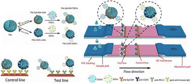
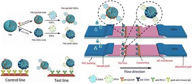
In recent years, the cargo profiles of extracellular vesicles (EVs), which were inherited from their parent cells, have emerged as a reliable biomarker for liquid biopsy (LB) in disease diagnosis, prognosis, and treatment monitoring. EVs secreted by different cells exhibit distinct characteristics, particularly in terms of disease diagnosis and prediction. However, currently available techniques for the quantitative analysis of EV cargoes, including enzyme-linked immunosorbent assay (ELISA), cannot specifically identify the cellular origin of EVs, thus seriously affecting the accuracy of EV-based liquid biopsy. In light of this, we here developed ultrabright fluorescent nanosphere (FNs)-based test strips which have the unique capability to specifically assess the levels of PD-L1-positive EVs (PD-L1+ EVs) derived from both tumor cells and immune cells in bodily fluids. The levels of PD-L1+ EV subpopulations in human saliva were quantified using the ultrabright fluorescent nanosphere-based test strips with more convenience and higher efficiency (detection time <30 min). Results demonstrated that the fluorescence intensity of the test line exhibited a good linear relationship respectively with the PD-L1 levels of tumor cell- (R2 = 0.993) and immune cell-derived EVs (R2 = 0.982) in human saliva. By assessing the levels of PD-L1+ EV subpopulations, our test strips hold immense potential for advancing the application of PD-L1+ EV subpopulation-based predictions in tumor diagnosis and prognosis evaluation. In summary, by integrating the benefits of FNs and lateral flow chromatography, we here provide a strategy to accurately measure the cargo levels of EVs originating from diverse cell sources in bodily fluids.
 求助内容:
求助内容: 应助结果提醒方式:


