{"title":"丝纤维水凝胶粘合剂在半月板撕裂修复中的新探索","authors":"Zhongwu Bei, Jing Zheng","doi":"10.1002/mba2.89","DOIUrl":null,"url":null,"abstract":"<p>Recently, in <i>Nature Communications</i>, Pan et al.<span><sup>1</sup></span> reported a novel dual-functional hydrogel bioadhesive (S-PIL10) based on silk fibroin, ionic liquid, and growth factor TGF-β1, achieving the seamless and dense reconstruction of torn meniscus. This kind of silk-based meniscus adhesive provides a revolutionary strategy for the repair of meniscal tears.</p><p>The meniscus, an essential elastic cartilaginous tissue within the knee joint, is between the femoral condyle and the tibial plateau, covering approximately two-thirds of the tibial surface.<span><sup>2</sup></span> It cushions impacts, distributes loads, maintains joint stability, and facilitates smooth joint motion. Meniscal injury is among the most prevalent musculoskeletal disorders affecting the knee, frequently arising from acute traumatic events, sports-related activities (such as sudden pivoting and stopping in basketball and soccer), or age-related degenerative alterations. Based on the pathological morphology of the injury, meniscal tears can be classified into vertical tears (longitudinal and radial tears), horizontal tears (most common), and complex tears (involving multiple tear patterns). These injuries may lead to debilitating symptoms, including pain, swelling, instability, and restricted mobility.<span><sup>3, 4</sup></span> Meniscal injuries primarily affect young individuals, characterized clinically by local bleeding, exudation, and acute inflammation. Left untreated, they may predispose individuals to early-onset osteoarthritis, significantly compromising their quality of life.</p><p>In clinical practice, incomplete meniscal tears without accompanying pathologies or small, stable peripheral tears may resolve without surgical intervention. However, the sparse distribution and poor vascularization of meniscal fibrocartilage cells, which occupy only 10%–30% of the meniscal thickness, often impede spontaneous healing, leading to the necessity of surgical intervention in most cases. Current treatment options primarily include meniscal repair, partial or complete meniscectomy, and allograft transplantation. Among these, meniscal repair aims to preserve as much healthy meniscal tissue as possible and is considered the gold standard in clinical practice. However, it is frequently constrained by tear location, size, and tissue quality. Conversely, meniscectomy addresses mechanical irritation from meniscal injury by removing the damaged portion or the entire meniscus. However, post-meniscectomy regeneration is limited, resulting in narrow, thin, and nonfunctional tissue. Although this approach can alleviate related symptoms, biomechanical studies indicate minimal meniscal tissue removal increases cartilage contact stress, reducing the natural meniscus's protective function. Furthermore, the overall failure rate of meniscal allograft transplantation is approximately 29% (4–14 years postoperatively), often accompanied by issues like joint space narrowing.</p><p>Tissue engineering and regenerative medicine technologies, incorporating scaffolds, cells, and biofactors, independently or in combination, offer promising avenues for treating meniscal injuries. While synthetic materials like polypropylene carbonate and polyethylene glycol have been used as repair scaffolds for meniscal and articular cartilage injuries, they may present challenges such as degradation-related toxicity, alterations in cell phenotype, and remodeling. The current research focus on meniscal repair materials primarily centers on: (1) optimizing biomaterials (biocompatibility and mechanical properties, etc.) to enhance the efficacy of meniscal repair; (2) utilizing stem cells and scaffold materials for meniscal tissue regeneration and repair; and (3) investigating emerging approaches such as growth factors and gene therapy to promote meniscal self-repair. However, in the transition to clinical application, there remain several key challenges. These include validating the safety and efficacy of new materials, material production and quality control, and ethical considerations. Overall, the clinical application of meniscal repair materials is still in continuous development and improvement, requiring multidisciplinary collaboration and long-term research investment.</p><p>Polymeric tissue adhesives typically provide mechanical support and hemostatic capabilities while sealing wound sites and preventing leakage, making them preferable to traditional surgical sutures and staples.<span><sup>5</sup></span> Due to their ease of use, minimal tissue trauma, and biological solid adhesion, tissue adhesives hold great promise in maintaining the integrity of natural meniscal tissue and ensuring tight adherence of damaged meniscal tissue.</p><p>Recently, Pan et al.<span><sup>1</sup></span> reported in the journal <i>Nature Communications</i> their study titled “Silk fibroin hydrogel adhesive enables sealed-tight reconstruction of meniscus tears.” Silk fibroin (SF) is a typical natural biomacromolecule with excellent biocompatibility and favorable characteristics for forming β-crystals, making it a promising biomaterial. Methacrylate-modified SF (SFMA) has been widely used in wound dressings, enzyme immobilization matrices, vascular grafts, and cartilage surface regeneration. The authors ingeniously combined SFMA, phenylboronic acid ionic liquid (PIL), and the growth factor TGF-β1 to obtain a hydrogel adhesive with outstanding performance (Figure 1). The PIL, custom-synthesized via alkylation reaction of 4-(bromomethyl) phenylboronic acid with 1-vinylimidazole, possesses three key characteristics: (1) the vinyl group in the imidazole cation serves as a comonomer; (2) the structure of the imidazole salt forms hydrogen bonds, promoting the formation of β-sheet structures for SF; and (3) the phenylboronic acid group reacts with hydroxyl groups in SF to form dynamic boronic ester bonds. Subsequently, the authors verified that incorporating PIL augmented the storage modulus and enhanced β-structures in the hydrogel adhesive, likely due to the suppression of polymer chain mobility within the hydrogel network and the Hoffmeister effect of PIL on SFMA. In a New Zealand rabbit model, the hydrogel adhesive S-PIL10 demonstrated significant efficacy in repairing meniscal tears and protecting cartilage from wear.</p><p>In summary, the authors employed a design strategy combining a biological adhesive with an ionic liquid to prepare the highly performing biological adhesive S-PIL10, which was successfully used to repair meniscal tears. The inaugural application of silk protein adhesive in meniscal tear repair holds significant revolutionary significance. (1) The clever utilization of SF's inherent β-crystalline characteristics to enhance mechanical performance design provides a new research avenue for developing other functional biomaterials based on material intrinsic property alterations (e.g., polyvinyl alcohol's crystallinity). (2) The application of biological adhesives may inspire more researchers to explore the applications of other biomaterials, thereby driving the development and innovation in the field of biological adhesives. (3) Compared to traditional synthetic materials, this biological adhesive exhibits superior environmental friendliness and degradability, offering guiding insights for developing safer and more effective biomaterials. (4) This study involves interdisciplinary collaboration across fields such as biomaterials science, chemistry, and biomedical engineering, and the successful application of S-PIL10 plays a positive role in promoting cooperation and communication among different disciplines. However, due to anatomical and physiological differences between rabbit and human menisci, further in-depth research is needed to evaluate its effectiveness in future clinical applications. We look forward to applying and validating this biological adhesive in chronic meniscal lesions, progressive tears, and large animal models, providing a solid research foundation for its feasibility in clinical applications. Additionally, we hope to see further optimization of its formulation and preparation processes to enhance its success rate in clinical applications.</p><p><b>Zhongwu Bei</b>: Conceptualization (equal); investigation (equal); writing—original draft (equal). <b>Jing Zheng</b>: Formal analysis (equal); supervision (equal); writing—review & editing (equal). All authors have read and approved the final manuscript.</p><p>The authors declare no conflict of interest.</p><p>Not applicable.</p>","PeriodicalId":100901,"journal":{"name":"MedComm – Biomaterials and Applications","volume":"3 3","pages":""},"PeriodicalIF":0.0000,"publicationDate":"2024-07-19","publicationTypes":"Journal Article","fieldsOfStudy":null,"isOpenAccess":false,"openAccessPdf":"https://onlinelibrary.wiley.com/doi/epdf/10.1002/mba2.89","citationCount":"0","resultStr":"{\"title\":\"Novel exploration of silk fibroin hydrogel adhesive in meniscal tear repair\",\"authors\":\"Zhongwu Bei, Jing Zheng\",\"doi\":\"10.1002/mba2.89\",\"DOIUrl\":null,\"url\":null,\"abstract\":\"<p>Recently, in <i>Nature Communications</i>, Pan et al.<span><sup>1</sup></span> reported a novel dual-functional hydrogel bioadhesive (S-PIL10) based on silk fibroin, ionic liquid, and growth factor TGF-β1, achieving the seamless and dense reconstruction of torn meniscus. This kind of silk-based meniscus adhesive provides a revolutionary strategy for the repair of meniscal tears.</p><p>The meniscus, an essential elastic cartilaginous tissue within the knee joint, is between the femoral condyle and the tibial plateau, covering approximately two-thirds of the tibial surface.<span><sup>2</sup></span> It cushions impacts, distributes loads, maintains joint stability, and facilitates smooth joint motion. Meniscal injury is among the most prevalent musculoskeletal disorders affecting the knee, frequently arising from acute traumatic events, sports-related activities (such as sudden pivoting and stopping in basketball and soccer), or age-related degenerative alterations. Based on the pathological morphology of the injury, meniscal tears can be classified into vertical tears (longitudinal and radial tears), horizontal tears (most common), and complex tears (involving multiple tear patterns). These injuries may lead to debilitating symptoms, including pain, swelling, instability, and restricted mobility.<span><sup>3, 4</sup></span> Meniscal injuries primarily affect young individuals, characterized clinically by local bleeding, exudation, and acute inflammation. Left untreated, they may predispose individuals to early-onset osteoarthritis, significantly compromising their quality of life.</p><p>In clinical practice, incomplete meniscal tears without accompanying pathologies or small, stable peripheral tears may resolve without surgical intervention. However, the sparse distribution and poor vascularization of meniscal fibrocartilage cells, which occupy only 10%–30% of the meniscal thickness, often impede spontaneous healing, leading to the necessity of surgical intervention in most cases. Current treatment options primarily include meniscal repair, partial or complete meniscectomy, and allograft transplantation. Among these, meniscal repair aims to preserve as much healthy meniscal tissue as possible and is considered the gold standard in clinical practice. However, it is frequently constrained by tear location, size, and tissue quality. Conversely, meniscectomy addresses mechanical irritation from meniscal injury by removing the damaged portion or the entire meniscus. However, post-meniscectomy regeneration is limited, resulting in narrow, thin, and nonfunctional tissue. Although this approach can alleviate related symptoms, biomechanical studies indicate minimal meniscal tissue removal increases cartilage contact stress, reducing the natural meniscus's protective function. Furthermore, the overall failure rate of meniscal allograft transplantation is approximately 29% (4–14 years postoperatively), often accompanied by issues like joint space narrowing.</p><p>Tissue engineering and regenerative medicine technologies, incorporating scaffolds, cells, and biofactors, independently or in combination, offer promising avenues for treating meniscal injuries. While synthetic materials like polypropylene carbonate and polyethylene glycol have been used as repair scaffolds for meniscal and articular cartilage injuries, they may present challenges such as degradation-related toxicity, alterations in cell phenotype, and remodeling. The current research focus on meniscal repair materials primarily centers on: (1) optimizing biomaterials (biocompatibility and mechanical properties, etc.) to enhance the efficacy of meniscal repair; (2) utilizing stem cells and scaffold materials for meniscal tissue regeneration and repair; and (3) investigating emerging approaches such as growth factors and gene therapy to promote meniscal self-repair. However, in the transition to clinical application, there remain several key challenges. These include validating the safety and efficacy of new materials, material production and quality control, and ethical considerations. Overall, the clinical application of meniscal repair materials is still in continuous development and improvement, requiring multidisciplinary collaboration and long-term research investment.</p><p>Polymeric tissue adhesives typically provide mechanical support and hemostatic capabilities while sealing wound sites and preventing leakage, making them preferable to traditional surgical sutures and staples.<span><sup>5</sup></span> Due to their ease of use, minimal tissue trauma, and biological solid adhesion, tissue adhesives hold great promise in maintaining the integrity of natural meniscal tissue and ensuring tight adherence of damaged meniscal tissue.</p><p>Recently, Pan et al.<span><sup>1</sup></span> reported in the journal <i>Nature Communications</i> their study titled “Silk fibroin hydrogel adhesive enables sealed-tight reconstruction of meniscus tears.” Silk fibroin (SF) is a typical natural biomacromolecule with excellent biocompatibility and favorable characteristics for forming β-crystals, making it a promising biomaterial. Methacrylate-modified SF (SFMA) has been widely used in wound dressings, enzyme immobilization matrices, vascular grafts, and cartilage surface regeneration. The authors ingeniously combined SFMA, phenylboronic acid ionic liquid (PIL), and the growth factor TGF-β1 to obtain a hydrogel adhesive with outstanding performance (Figure 1). The PIL, custom-synthesized via alkylation reaction of 4-(bromomethyl) phenylboronic acid with 1-vinylimidazole, possesses three key characteristics: (1) the vinyl group in the imidazole cation serves as a comonomer; (2) the structure of the imidazole salt forms hydrogen bonds, promoting the formation of β-sheet structures for SF; and (3) the phenylboronic acid group reacts with hydroxyl groups in SF to form dynamic boronic ester bonds. Subsequently, the authors verified that incorporating PIL augmented the storage modulus and enhanced β-structures in the hydrogel adhesive, likely due to the suppression of polymer chain mobility within the hydrogel network and the Hoffmeister effect of PIL on SFMA. In a New Zealand rabbit model, the hydrogel adhesive S-PIL10 demonstrated significant efficacy in repairing meniscal tears and protecting cartilage from wear.</p><p>In summary, the authors employed a design strategy combining a biological adhesive with an ionic liquid to prepare the highly performing biological adhesive S-PIL10, which was successfully used to repair meniscal tears. The inaugural application of silk protein adhesive in meniscal tear repair holds significant revolutionary significance. (1) The clever utilization of SF's inherent β-crystalline characteristics to enhance mechanical performance design provides a new research avenue for developing other functional biomaterials based on material intrinsic property alterations (e.g., polyvinyl alcohol's crystallinity). (2) The application of biological adhesives may inspire more researchers to explore the applications of other biomaterials, thereby driving the development and innovation in the field of biological adhesives. (3) Compared to traditional synthetic materials, this biological adhesive exhibits superior environmental friendliness and degradability, offering guiding insights for developing safer and more effective biomaterials. (4) This study involves interdisciplinary collaboration across fields such as biomaterials science, chemistry, and biomedical engineering, and the successful application of S-PIL10 plays a positive role in promoting cooperation and communication among different disciplines. However, due to anatomical and physiological differences between rabbit and human menisci, further in-depth research is needed to evaluate its effectiveness in future clinical applications. We look forward to applying and validating this biological adhesive in chronic meniscal lesions, progressive tears, and large animal models, providing a solid research foundation for its feasibility in clinical applications. Additionally, we hope to see further optimization of its formulation and preparation processes to enhance its success rate in clinical applications.</p><p><b>Zhongwu Bei</b>: Conceptualization (equal); investigation (equal); writing—original draft (equal). <b>Jing Zheng</b>: Formal analysis (equal); supervision (equal); writing—review & editing (equal). All authors have read and approved the final manuscript.</p><p>The authors declare no conflict of interest.</p><p>Not applicable.</p>\",\"PeriodicalId\":100901,\"journal\":{\"name\":\"MedComm – Biomaterials and Applications\",\"volume\":\"3 3\",\"pages\":\"\"},\"PeriodicalIF\":0.0000,\"publicationDate\":\"2024-07-19\",\"publicationTypes\":\"Journal Article\",\"fieldsOfStudy\":null,\"isOpenAccess\":false,\"openAccessPdf\":\"https://onlinelibrary.wiley.com/doi/epdf/10.1002/mba2.89\",\"citationCount\":\"0\",\"resultStr\":null,\"platform\":\"Semanticscholar\",\"paperid\":null,\"PeriodicalName\":\"MedComm – Biomaterials and Applications\",\"FirstCategoryId\":\"1085\",\"ListUrlMain\":\"https://onlinelibrary.wiley.com/doi/10.1002/mba2.89\",\"RegionNum\":0,\"RegionCategory\":null,\"ArticlePicture\":[],\"TitleCN\":null,\"AbstractTextCN\":null,\"PMCID\":null,\"EPubDate\":\"\",\"PubModel\":\"\",\"JCR\":\"\",\"JCRName\":\"\",\"Score\":null,\"Total\":0}","platform":"Semanticscholar","paperid":null,"PeriodicalName":"MedComm – Biomaterials and Applications","FirstCategoryId":"1085","ListUrlMain":"https://onlinelibrary.wiley.com/doi/10.1002/mba2.89","RegionNum":0,"RegionCategory":null,"ArticlePicture":[],"TitleCN":null,"AbstractTextCN":null,"PMCID":null,"EPubDate":"","PubModel":"","JCR":"","JCRName":"","Score":null,"Total":0}
Novel exploration of silk fibroin hydrogel adhesive in meniscal tear repair
Recently, in Nature Communications, Pan et al.1 reported a novel dual-functional hydrogel bioadhesive (S-PIL10) based on silk fibroin, ionic liquid, and growth factor TGF-β1, achieving the seamless and dense reconstruction of torn meniscus. This kind of silk-based meniscus adhesive provides a revolutionary strategy for the repair of meniscal tears.
The meniscus, an essential elastic cartilaginous tissue within the knee joint, is between the femoral condyle and the tibial plateau, covering approximately two-thirds of the tibial surface.2 It cushions impacts, distributes loads, maintains joint stability, and facilitates smooth joint motion. Meniscal injury is among the most prevalent musculoskeletal disorders affecting the knee, frequently arising from acute traumatic events, sports-related activities (such as sudden pivoting and stopping in basketball and soccer), or age-related degenerative alterations. Based on the pathological morphology of the injury, meniscal tears can be classified into vertical tears (longitudinal and radial tears), horizontal tears (most common), and complex tears (involving multiple tear patterns). These injuries may lead to debilitating symptoms, including pain, swelling, instability, and restricted mobility.3, 4 Meniscal injuries primarily affect young individuals, characterized clinically by local bleeding, exudation, and acute inflammation. Left untreated, they may predispose individuals to early-onset osteoarthritis, significantly compromising their quality of life.
In clinical practice, incomplete meniscal tears without accompanying pathologies or small, stable peripheral tears may resolve without surgical intervention. However, the sparse distribution and poor vascularization of meniscal fibrocartilage cells, which occupy only 10%–30% of the meniscal thickness, often impede spontaneous healing, leading to the necessity of surgical intervention in most cases. Current treatment options primarily include meniscal repair, partial or complete meniscectomy, and allograft transplantation. Among these, meniscal repair aims to preserve as much healthy meniscal tissue as possible and is considered the gold standard in clinical practice. However, it is frequently constrained by tear location, size, and tissue quality. Conversely, meniscectomy addresses mechanical irritation from meniscal injury by removing the damaged portion or the entire meniscus. However, post-meniscectomy regeneration is limited, resulting in narrow, thin, and nonfunctional tissue. Although this approach can alleviate related symptoms, biomechanical studies indicate minimal meniscal tissue removal increases cartilage contact stress, reducing the natural meniscus's protective function. Furthermore, the overall failure rate of meniscal allograft transplantation is approximately 29% (4–14 years postoperatively), often accompanied by issues like joint space narrowing.
Tissue engineering and regenerative medicine technologies, incorporating scaffolds, cells, and biofactors, independently or in combination, offer promising avenues for treating meniscal injuries. While synthetic materials like polypropylene carbonate and polyethylene glycol have been used as repair scaffolds for meniscal and articular cartilage injuries, they may present challenges such as degradation-related toxicity, alterations in cell phenotype, and remodeling. The current research focus on meniscal repair materials primarily centers on: (1) optimizing biomaterials (biocompatibility and mechanical properties, etc.) to enhance the efficacy of meniscal repair; (2) utilizing stem cells and scaffold materials for meniscal tissue regeneration and repair; and (3) investigating emerging approaches such as growth factors and gene therapy to promote meniscal self-repair. However, in the transition to clinical application, there remain several key challenges. These include validating the safety and efficacy of new materials, material production and quality control, and ethical considerations. Overall, the clinical application of meniscal repair materials is still in continuous development and improvement, requiring multidisciplinary collaboration and long-term research investment.
Polymeric tissue adhesives typically provide mechanical support and hemostatic capabilities while sealing wound sites and preventing leakage, making them preferable to traditional surgical sutures and staples.5 Due to their ease of use, minimal tissue trauma, and biological solid adhesion, tissue adhesives hold great promise in maintaining the integrity of natural meniscal tissue and ensuring tight adherence of damaged meniscal tissue.
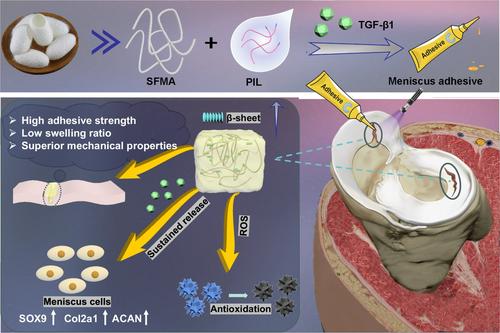
Recently, Pan et al.1 reported in the journal Nature Communications their study titled “Silk fibroin hydrogel adhesive enables sealed-tight reconstruction of meniscus tears.” Silk fibroin (SF) is a typical natural biomacromolecule with excellent biocompatibility and favorable characteristics for forming β-crystals, making it a promising biomaterial. Methacrylate-modified SF (SFMA) has been widely used in wound dressings, enzyme immobilization matrices, vascular grafts, and cartilage surface regeneration. The authors ingeniously combined SFMA, phenylboronic acid ionic liquid (PIL), and the growth factor TGF-β1 to obtain a hydrogel adhesive with outstanding performance (Figure 1). The PIL, custom-synthesized via alkylation reaction of 4-(bromomethyl) phenylboronic acid with 1-vinylimidazole, possesses three key characteristics: (1) the vinyl group in the imidazole cation serves as a comonomer; (2) the structure of the imidazole salt forms hydrogen bonds, promoting the formation of β-sheet structures for SF; and (3) the phenylboronic acid group reacts with hydroxyl groups in SF to form dynamic boronic ester bonds. Subsequently, the authors verified that incorporating PIL augmented the storage modulus and enhanced β-structures in the hydrogel adhesive, likely due to the suppression of polymer chain mobility within the hydrogel network and the Hoffmeister effect of PIL on SFMA. In a New Zealand rabbit model, the hydrogel adhesive S-PIL10 demonstrated significant efficacy in repairing meniscal tears and protecting cartilage from wear.
In summary, the authors employed a design strategy combining a biological adhesive with an ionic liquid to prepare the highly performing biological adhesive S-PIL10, which was successfully used to repair meniscal tears. The inaugural application of silk protein adhesive in meniscal tear repair holds significant revolutionary significance. (1) The clever utilization of SF's inherent β-crystalline characteristics to enhance mechanical performance design provides a new research avenue for developing other functional biomaterials based on material intrinsic property alterations (e.g., polyvinyl alcohol's crystallinity). (2) The application of biological adhesives may inspire more researchers to explore the applications of other biomaterials, thereby driving the development and innovation in the field of biological adhesives. (3) Compared to traditional synthetic materials, this biological adhesive exhibits superior environmental friendliness and degradability, offering guiding insights for developing safer and more effective biomaterials. (4) This study involves interdisciplinary collaboration across fields such as biomaterials science, chemistry, and biomedical engineering, and the successful application of S-PIL10 plays a positive role in promoting cooperation and communication among different disciplines. However, due to anatomical and physiological differences between rabbit and human menisci, further in-depth research is needed to evaluate its effectiveness in future clinical applications. We look forward to applying and validating this biological adhesive in chronic meniscal lesions, progressive tears, and large animal models, providing a solid research foundation for its feasibility in clinical applications. Additionally, we hope to see further optimization of its formulation and preparation processes to enhance its success rate in clinical applications.
Zhongwu Bei: Conceptualization (equal); investigation (equal); writing—original draft (equal). Jing Zheng: Formal analysis (equal); supervision (equal); writing—review & editing (equal). All authors have read and approved the final manuscript.

 求助内容:
求助内容: 应助结果提醒方式:


