Petra Poláčková, Jiří Borovec, Jana Vašáková, Matěj Patzelt, Wanda Urbanová, Michaela Mihulová, Milan Macek Jr, Markéta Havlovicová, Veronika Moslerová
{"title":"使用三维几何形态测量法对眼耳鼻椎体频谱患者的面部进行分析。","authors":"Petra Poláčková, Jiří Borovec, Jana Vašáková, Matěj Patzelt, Wanda Urbanová, Michaela Mihulová, Milan Macek Jr, Markéta Havlovicová, Veronika Moslerová","doi":"10.1111/ocr.12834","DOIUrl":null,"url":null,"abstract":"<div>\n \n \n <section>\n \n <h3> Aim</h3>\n \n <p>To utilize three-dimensional (3D) geometric morphometry for visualization of the level of facial asymmetry in patients with the oculo-auriculo-vertebral spectrum (OAVS).</p>\n </section>\n \n <section>\n \n <h3> Materials and Methods</h3>\n \n <p>Three-dimensional facial scans of 25 Czech patients with OAVS were processed. The patients were divided into subgroups according to Pruzansky classification. For 13 of them, second 3D facial scans were obtained. The 3D facial scans were processed using geometric morphometry. Soft tissue facial asymmetry in the sagittal plane and its changes in two time spots were visualized using colour-coded maps with a thermometre-like scale.</p>\n </section>\n \n <section>\n \n <h3> Results</h3>\n \n <p>Individual facial asymmetry was visualized in all patients as well as the mean facial asymmetry for every Pruzansky subgroup. The mean colour-coded maps of type I and type IIA subgroups showed no differences in facial asymmetry, more pronounced asymmetry in the middle and the lower facial third was found between type IIA and type IIB (maximum 1.5 mm) and between type IIB and type III (maximum 2 mm). The degree of intensity facial asymmetry in affected middle and lower facial thirds did not change distinctly during the two time spots in all subgroups.</p>\n </section>\n \n <section>\n \n <h3> Conclusions</h3>\n \n <p>The 3D geometric morphometry in OAVS patients could be a useful tool for objective facial asymmetry assessment in patients with OAVS. The calculated colour-coded maps are illustrative and useful for clinical evaluation.</p>\n </section>\n </div>","PeriodicalId":19652,"journal":{"name":"Orthodontics & Craniofacial Research","volume":"27 6","pages":"917-927"},"PeriodicalIF":2.4000,"publicationDate":"2024-07-19","publicationTypes":"Journal Article","fieldsOfStudy":null,"isOpenAccess":false,"openAccessPdf":"https://onlinelibrary.wiley.com/doi/epdf/10.1111/ocr.12834","citationCount":"0","resultStr":"{\"title\":\"Using three-dimensional geometric morphometry for facial analysis in patients with the oculo-auriculo-vertebral spectrum\",\"authors\":\"Petra Poláčková, Jiří Borovec, Jana Vašáková, Matěj Patzelt, Wanda Urbanová, Michaela Mihulová, Milan Macek Jr, Markéta Havlovicová, Veronika Moslerová\",\"doi\":\"10.1111/ocr.12834\",\"DOIUrl\":null,\"url\":null,\"abstract\":\"<div>\\n \\n \\n <section>\\n \\n <h3> Aim</h3>\\n \\n <p>To utilize three-dimensional (3D) geometric morphometry for visualization of the level of facial asymmetry in patients with the oculo-auriculo-vertebral spectrum (OAVS).</p>\\n </section>\\n \\n <section>\\n \\n <h3> Materials and Methods</h3>\\n \\n <p>Three-dimensional facial scans of 25 Czech patients with OAVS were processed. The patients were divided into subgroups according to Pruzansky classification. For 13 of them, second 3D facial scans were obtained. The 3D facial scans were processed using geometric morphometry. Soft tissue facial asymmetry in the sagittal plane and its changes in two time spots were visualized using colour-coded maps with a thermometre-like scale.</p>\\n </section>\\n \\n <section>\\n \\n <h3> Results</h3>\\n \\n <p>Individual facial asymmetry was visualized in all patients as well as the mean facial asymmetry for every Pruzansky subgroup. The mean colour-coded maps of type I and type IIA subgroups showed no differences in facial asymmetry, more pronounced asymmetry in the middle and the lower facial third was found between type IIA and type IIB (maximum 1.5 mm) and between type IIB and type III (maximum 2 mm). The degree of intensity facial asymmetry in affected middle and lower facial thirds did not change distinctly during the two time spots in all subgroups.</p>\\n </section>\\n \\n <section>\\n \\n <h3> Conclusions</h3>\\n \\n <p>The 3D geometric morphometry in OAVS patients could be a useful tool for objective facial asymmetry assessment in patients with OAVS. The calculated colour-coded maps are illustrative and useful for clinical evaluation.</p>\\n </section>\\n </div>\",\"PeriodicalId\":19652,\"journal\":{\"name\":\"Orthodontics & Craniofacial Research\",\"volume\":\"27 6\",\"pages\":\"917-927\"},\"PeriodicalIF\":2.4000,\"publicationDate\":\"2024-07-19\",\"publicationTypes\":\"Journal Article\",\"fieldsOfStudy\":null,\"isOpenAccess\":false,\"openAccessPdf\":\"https://onlinelibrary.wiley.com/doi/epdf/10.1111/ocr.12834\",\"citationCount\":\"0\",\"resultStr\":null,\"platform\":\"Semanticscholar\",\"paperid\":null,\"PeriodicalName\":\"Orthodontics & Craniofacial Research\",\"FirstCategoryId\":\"3\",\"ListUrlMain\":\"https://onlinelibrary.wiley.com/doi/10.1111/ocr.12834\",\"RegionNum\":3,\"RegionCategory\":\"医学\",\"ArticlePicture\":[],\"TitleCN\":null,\"AbstractTextCN\":null,\"PMCID\":null,\"EPubDate\":\"\",\"PubModel\":\"\",\"JCR\":\"Q2\",\"JCRName\":\"DENTISTRY, ORAL SURGERY & MEDICINE\",\"Score\":null,\"Total\":0}","platform":"Semanticscholar","paperid":null,"PeriodicalName":"Orthodontics & Craniofacial Research","FirstCategoryId":"3","ListUrlMain":"https://onlinelibrary.wiley.com/doi/10.1111/ocr.12834","RegionNum":3,"RegionCategory":"医学","ArticlePicture":[],"TitleCN":null,"AbstractTextCN":null,"PMCID":null,"EPubDate":"","PubModel":"","JCR":"Q2","JCRName":"DENTISTRY, ORAL SURGERY & MEDICINE","Score":null,"Total":0}
Using three-dimensional geometric morphometry for facial analysis in patients with the oculo-auriculo-vertebral spectrum
Aim
To utilize three-dimensional (3D) geometric morphometry for visualization of the level of facial asymmetry in patients with the oculo-auriculo-vertebral spectrum (OAVS).
Materials and Methods
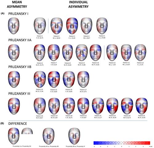
Three-dimensional facial scans of 25 Czech patients with OAVS were processed. The patients were divided into subgroups according to Pruzansky classification. For 13 of them, second 3D facial scans were obtained. The 3D facial scans were processed using geometric morphometry. Soft tissue facial asymmetry in the sagittal plane and its changes in two time spots were visualized using colour-coded maps with a thermometre-like scale.
Results
Individual facial asymmetry was visualized in all patients as well as the mean facial asymmetry for every Pruzansky subgroup. The mean colour-coded maps of type I and type IIA subgroups showed no differences in facial asymmetry, more pronounced asymmetry in the middle and the lower facial third was found between type IIA and type IIB (maximum 1.5 mm) and between type IIB and type III (maximum 2 mm). The degree of intensity facial asymmetry in affected middle and lower facial thirds did not change distinctly during the two time spots in all subgroups.
Conclusions
The 3D geometric morphometry in OAVS patients could be a useful tool for objective facial asymmetry assessment in patients with OAVS. The calculated colour-coded maps are illustrative and useful for clinical evaluation.
期刊介绍:
Orthodontics & Craniofacial Research - Genes, Growth and Development is published to serve its readers as an international forum for the presentation and critical discussion of issues pertinent to the advancement of the specialty of orthodontics and the evidence-based knowledge of craniofacial growth and development. This forum is based on scientifically supported information, but also includes minority and conflicting opinions.
The objective of the journal is to facilitate effective communication between the research community and practicing clinicians. Original papers of high scientific quality that report the findings of clinical trials, clinical epidemiology, and novel therapeutic or diagnostic approaches are appropriate submissions. Similarly, we welcome papers in genetics, developmental biology, syndromology, surgery, speech and hearing, and other biomedical disciplines related to clinical orthodontics and normal and abnormal craniofacial growth and development. In addition to original and basic research, the journal publishes concise reviews, case reports of substantial value, invited essays, letters, and announcements.
The journal is published quarterly. The review of submitted papers will be coordinated by the editor and members of the editorial board. It is policy to review manuscripts within 3 to 4 weeks of receipt and to publish within 3 to 6 months of acceptance.
 求助内容:
求助内容: 应助结果提醒方式:


