Ji Hyun Kim, Satoshi Ishizuka, Kei Kitamura, Gen Murakami, José Francisco Rodríguez-Vázquez, Shin-ichi Abe, Masataka Kasahara
{"title":"踝关节从最初的小关节和距骨内外侧排列的本体转化:对人类胚胎和早期胎儿的组织学研究。","authors":"Ji Hyun Kim, Satoshi Ishizuka, Kei Kitamura, Gen Murakami, José Francisco Rodríguez-Vázquez, Shin-ichi Abe, Masataka Kasahara","doi":"10.1111/joa.14039","DOIUrl":null,"url":null,"abstract":"<p>The human calcaneus is robust and provides a prominent heel for effective bipedal locomotion, although the adjacent talus has no muscle attachments. However, there is incomplete information about the morphological changes in these prominent bones during embryo development. We examined serial histological sections of 23 human embryos and early-term fetuses (approximately 5–10 weeks' gestational age [GA]). At a GA of 5 weeks, the precartilage talus was parallel to and on the medial side of the calcaneus, which had a prolate spheroid shape and consisted of three masses. At a GA of 6 weeks, the cartilaginous talus extended along the proximodistal axis, and the tuber calcanei became long and bulky, with a small sustentaculum talus at the “distal” side. At a GA of 6 to 8 weeks, the sustentaculum had a medial extension below the talus so that the talus “rode over” the calcaneus. In contrast, the talus had a more complex shape, depending on the growth of adjacent bones. At a GA of 9 to 10 weeks, the talus was above the calcaneus, but the medial part still faced the plantar subcutaneous tissue because of the relatively small sustentaculum. Therefore, the final morphology appeared after an additional several weeks. Muscle activity seemed to facilitate growth of the tuber calcanei, but growth of the other parts of calcaneus, including the sustentaculum, seemed to depend on active proliferation at the different sites of cartilage. Multiple tendons and ligaments seemed to fix the talus so that it remained close to the calcaneus.</p>","PeriodicalId":14971,"journal":{"name":"Journal of Anatomy","volume":null,"pages":null},"PeriodicalIF":1.8000,"publicationDate":"2024-07-20","publicationTypes":"Journal Article","fieldsOfStudy":null,"isOpenAccess":false,"openAccessPdf":"","citationCount":"0","resultStr":"{\"title\":\"Ontogenic transformation of the ankle from the initial mediolateral arrangement of the calcaneus and talus: A histological study of human embryos and early fetuses\",\"authors\":\"Ji Hyun Kim, Satoshi Ishizuka, Kei Kitamura, Gen Murakami, José Francisco Rodríguez-Vázquez, Shin-ichi Abe, Masataka Kasahara\",\"doi\":\"10.1111/joa.14039\",\"DOIUrl\":null,\"url\":null,\"abstract\":\"<p>The human calcaneus is robust and provides a prominent heel for effective bipedal locomotion, although the adjacent talus has no muscle attachments. However, there is incomplete information about the morphological changes in these prominent bones during embryo development. We examined serial histological sections of 23 human embryos and early-term fetuses (approximately 5–10 weeks' gestational age [GA]). At a GA of 5 weeks, the precartilage talus was parallel to and on the medial side of the calcaneus, which had a prolate spheroid shape and consisted of three masses. At a GA of 6 weeks, the cartilaginous talus extended along the proximodistal axis, and the tuber calcanei became long and bulky, with a small sustentaculum talus at the “distal” side. At a GA of 6 to 8 weeks, the sustentaculum had a medial extension below the talus so that the talus “rode over” the calcaneus. In contrast, the talus had a more complex shape, depending on the growth of adjacent bones. At a GA of 9 to 10 weeks, the talus was above the calcaneus, but the medial part still faced the plantar subcutaneous tissue because of the relatively small sustentaculum. Therefore, the final morphology appeared after an additional several weeks. Muscle activity seemed to facilitate growth of the tuber calcanei, but growth of the other parts of calcaneus, including the sustentaculum, seemed to depend on active proliferation at the different sites of cartilage. Multiple tendons and ligaments seemed to fix the talus so that it remained close to the calcaneus.</p>\",\"PeriodicalId\":14971,\"journal\":{\"name\":\"Journal of Anatomy\",\"volume\":null,\"pages\":null},\"PeriodicalIF\":1.8000,\"publicationDate\":\"2024-07-20\",\"publicationTypes\":\"Journal Article\",\"fieldsOfStudy\":null,\"isOpenAccess\":false,\"openAccessPdf\":\"\",\"citationCount\":\"0\",\"resultStr\":null,\"platform\":\"Semanticscholar\",\"paperid\":null,\"PeriodicalName\":\"Journal of Anatomy\",\"FirstCategoryId\":\"3\",\"ListUrlMain\":\"https://onlinelibrary.wiley.com/doi/10.1111/joa.14039\",\"RegionNum\":3,\"RegionCategory\":\"医学\",\"ArticlePicture\":[],\"TitleCN\":null,\"AbstractTextCN\":null,\"PMCID\":null,\"EPubDate\":\"\",\"PubModel\":\"\",\"JCR\":\"Q2\",\"JCRName\":\"ANATOMY & MORPHOLOGY\",\"Score\":null,\"Total\":0}","platform":"Semanticscholar","paperid":null,"PeriodicalName":"Journal of Anatomy","FirstCategoryId":"3","ListUrlMain":"https://onlinelibrary.wiley.com/doi/10.1111/joa.14039","RegionNum":3,"RegionCategory":"医学","ArticlePicture":[],"TitleCN":null,"AbstractTextCN":null,"PMCID":null,"EPubDate":"","PubModel":"","JCR":"Q2","JCRName":"ANATOMY & MORPHOLOGY","Score":null,"Total":0}
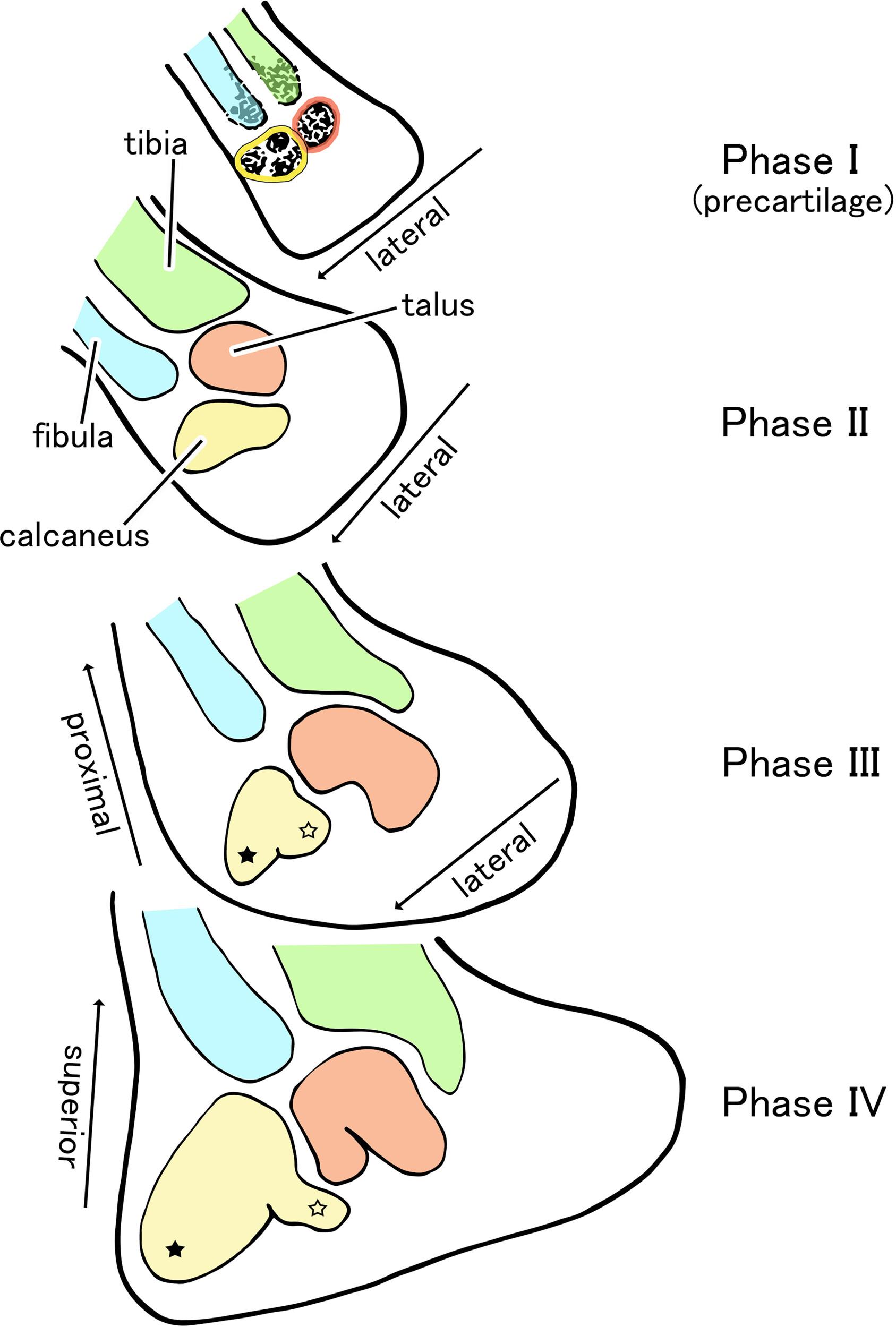
Ontogenic transformation of the ankle from the initial mediolateral arrangement of the calcaneus and talus: A histological study of human embryos and early fetuses
The human calcaneus is robust and provides a prominent heel for effective bipedal locomotion, although the adjacent talus has no muscle attachments. However, there is incomplete information about the morphological changes in these prominent bones during embryo development. We examined serial histological sections of 23 human embryos and early-term fetuses (approximately 5–10 weeks' gestational age [GA]). At a GA of 5 weeks, the precartilage talus was parallel to and on the medial side of the calcaneus, which had a prolate spheroid shape and consisted of three masses. At a GA of 6 weeks, the cartilaginous talus extended along the proximodistal axis, and the tuber calcanei became long and bulky, with a small sustentaculum talus at the “distal” side. At a GA of 6 to 8 weeks, the sustentaculum had a medial extension below the talus so that the talus “rode over” the calcaneus. In contrast, the talus had a more complex shape, depending on the growth of adjacent bones. At a GA of 9 to 10 weeks, the talus was above the calcaneus, but the medial part still faced the plantar subcutaneous tissue because of the relatively small sustentaculum. Therefore, the final morphology appeared after an additional several weeks. Muscle activity seemed to facilitate growth of the tuber calcanei, but growth of the other parts of calcaneus, including the sustentaculum, seemed to depend on active proliferation at the different sites of cartilage. Multiple tendons and ligaments seemed to fix the talus so that it remained close to the calcaneus.
期刊介绍:
Journal of Anatomy is an international peer-reviewed journal sponsored by the Anatomical Society. The journal publishes original papers, invited review articles and book reviews. Its main focus is to understand anatomy through an analysis of structure, function, development and evolution. Priority will be given to studies of that clearly articulate their relevance to the anatomical community. Focal areas include: experimental studies, contributions based on molecular and cell biology and on the application of modern imaging techniques and papers with novel methods or synthetic perspective on an anatomical system.
Studies that are essentially descriptive anatomy are appropriate only if they communicate clearly a broader functional or evolutionary significance. You must clearly state the broader implications of your work in the abstract.
We particularly welcome submissions in the following areas:
Cell biology and tissue architecture
Comparative functional morphology
Developmental biology
Evolutionary developmental biology
Evolutionary morphology
Functional human anatomy
Integrative vertebrate paleontology
Methodological innovations in anatomical research
Musculoskeletal system
Neuroanatomy and neurodegeneration
Significant advances in anatomical education.

 求助内容:
求助内容: 应助结果提醒方式:


