Lorenz Jacob Mangahas, Rowena Wea Reyes, Richmond Siazon
{"title":"菲律宾青年原发性结膜基底细胞癌模仿眼表鳞状上皮细胞瘤:病例报告和文献综述。","authors":"Lorenz Jacob Mangahas, Rowena Wea Reyes, Richmond Siazon","doi":"10.1155/2024/3113342","DOIUrl":null,"url":null,"abstract":"<p><p><b>Objective:</b> To describe the morphological and histopathological features of primary conjunctival basal cell carcinoma (BCC) in a young adult Filipino. <b>Introduction:</b> Malignant conjunctival tumors arise from different cells, the most common of which are squamous cell carcinomas (SCCs), (including ocular surface squamous neoplasia [OSSN]), melanomas, and lymphomas. Primary conjunctival BCC is rare and can mimic the clinical features of OSSN. Only seven reported cases were published. Most cases are in the 6th-8th decades of life, and we report the first case in a young adult male. <b>Case Summary:</b> A 37/M, HIV-seronegative, presenting with a 3-year history of enlarging fleshy, pedunculated mass on the right eye measuring 8.5 mm × 8.0 mm at the superonasal limbus encroaching on the cornea, with prominent feeder vessels. Whitish-to-grayish plaques were observed on the surface of the lesions. Wide excision of the mass using the no-touch technique was performed under local anesthesia. Four cycles of mitomycin C (0.02%) were administered as chemoadjuvant therapy. Histopathology showed basaloid cells with peripheral palisading, most consistent with BCC. Immunohistochemistry was positive for Bcl-2 and CD10 markers and negative for epithelial membrane antigen (EMA) and carcinoembryonic antigen (CEA), confirming conjunctival BCC. Eight weeks postoperatively, fibrovascular tissue proliferation was noted at the excision site. Anterior segment-optical coherence tomography (AS-OCT) revealed a thickened hyperreflective band that was continuous with the epithelium, indicating possible recurrence. Resection with rush frozen section revealed fibrotic tissue that was negative for tumor cells. The bare sclera was covered with conjunctival autograft. There was no recurrence of the lesion after 16 months of follow-up. <b>Conclusion:</b> Primary BCC of the conjunctiva is rarely encountered, especially in young individuals, mimicking squamous neoplasia both in morphology and histopathology. Therefore, this should be considered in the differential diagnosis of OSSN. Immunostaining is crucial in differentiating between the two conditions and confirming the diagnosis. In most cases, wide surgical excision is sufficient. In addition, adjuvant therapies may be beneficial in preventing tumor recurrence.</p>","PeriodicalId":9603,"journal":{"name":"Case Reports in Ophthalmological Medicine","volume":"2024 ","pages":"3113342"},"PeriodicalIF":0.4000,"publicationDate":"2024-07-11","publicationTypes":"Journal Article","fieldsOfStudy":null,"isOpenAccess":false,"openAccessPdf":"https://www.ncbi.nlm.nih.gov/pmc/articles/PMC11257755/pdf/","citationCount":"0","resultStr":"{\"title\":\"Primary Conjunctival Basal Cell Carcinoma Mimicking an Ocular Surface Squamous Neoplasia in a Young Adult Filipino: A Case Report and Literature Review.\",\"authors\":\"Lorenz Jacob Mangahas, Rowena Wea Reyes, Richmond Siazon\",\"doi\":\"10.1155/2024/3113342\",\"DOIUrl\":null,\"url\":null,\"abstract\":\"<p><p><b>Objective:</b> To describe the morphological and histopathological features of primary conjunctival basal cell carcinoma (BCC) in a young adult Filipino. <b>Introduction:</b> Malignant conjunctival tumors arise from different cells, the most common of which are squamous cell carcinomas (SCCs), (including ocular surface squamous neoplasia [OSSN]), melanomas, and lymphomas. Primary conjunctival BCC is rare and can mimic the clinical features of OSSN. Only seven reported cases were published. Most cases are in the 6th-8th decades of life, and we report the first case in a young adult male. <b>Case Summary:</b> A 37/M, HIV-seronegative, presenting with a 3-year history of enlarging fleshy, pedunculated mass on the right eye measuring 8.5 mm × 8.0 mm at the superonasal limbus encroaching on the cornea, with prominent feeder vessels. Whitish-to-grayish plaques were observed on the surface of the lesions. Wide excision of the mass using the no-touch technique was performed under local anesthesia. Four cycles of mitomycin C (0.02%) were administered as chemoadjuvant therapy. Histopathology showed basaloid cells with peripheral palisading, most consistent with BCC. Immunohistochemistry was positive for Bcl-2 and CD10 markers and negative for epithelial membrane antigen (EMA) and carcinoembryonic antigen (CEA), confirming conjunctival BCC. Eight weeks postoperatively, fibrovascular tissue proliferation was noted at the excision site. Anterior segment-optical coherence tomography (AS-OCT) revealed a thickened hyperreflective band that was continuous with the epithelium, indicating possible recurrence. Resection with rush frozen section revealed fibrotic tissue that was negative for tumor cells. The bare sclera was covered with conjunctival autograft. There was no recurrence of the lesion after 16 months of follow-up. <b>Conclusion:</b> Primary BCC of the conjunctiva is rarely encountered, especially in young individuals, mimicking squamous neoplasia both in morphology and histopathology. Therefore, this should be considered in the differential diagnosis of OSSN. Immunostaining is crucial in differentiating between the two conditions and confirming the diagnosis. In most cases, wide surgical excision is sufficient. In addition, adjuvant therapies may be beneficial in preventing tumor recurrence.</p>\",\"PeriodicalId\":9603,\"journal\":{\"name\":\"Case Reports in Ophthalmological Medicine\",\"volume\":\"2024 \",\"pages\":\"3113342\"},\"PeriodicalIF\":0.4000,\"publicationDate\":\"2024-07-11\",\"publicationTypes\":\"Journal Article\",\"fieldsOfStudy\":null,\"isOpenAccess\":false,\"openAccessPdf\":\"https://www.ncbi.nlm.nih.gov/pmc/articles/PMC11257755/pdf/\",\"citationCount\":\"0\",\"resultStr\":null,\"platform\":\"Semanticscholar\",\"paperid\":null,\"PeriodicalName\":\"Case Reports in Ophthalmological Medicine\",\"FirstCategoryId\":\"1085\",\"ListUrlMain\":\"https://doi.org/10.1155/2024/3113342\",\"RegionNum\":0,\"RegionCategory\":null,\"ArticlePicture\":[],\"TitleCN\":null,\"AbstractTextCN\":null,\"PMCID\":null,\"EPubDate\":\"2024/1/1 0:00:00\",\"PubModel\":\"eCollection\",\"JCR\":\"Q4\",\"JCRName\":\"OPHTHALMOLOGY\",\"Score\":null,\"Total\":0}","platform":"Semanticscholar","paperid":null,"PeriodicalName":"Case Reports in Ophthalmological Medicine","FirstCategoryId":"1085","ListUrlMain":"https://doi.org/10.1155/2024/3113342","RegionNum":0,"RegionCategory":null,"ArticlePicture":[],"TitleCN":null,"AbstractTextCN":null,"PMCID":null,"EPubDate":"2024/1/1 0:00:00","PubModel":"eCollection","JCR":"Q4","JCRName":"OPHTHALMOLOGY","Score":null,"Total":0}
Primary Conjunctival Basal Cell Carcinoma Mimicking an Ocular Surface Squamous Neoplasia in a Young Adult Filipino: A Case Report and Literature Review.
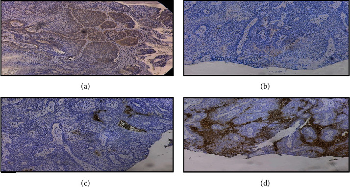
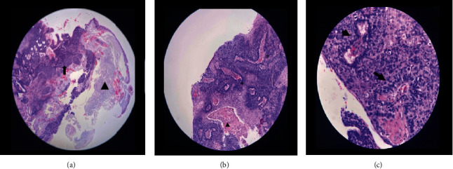
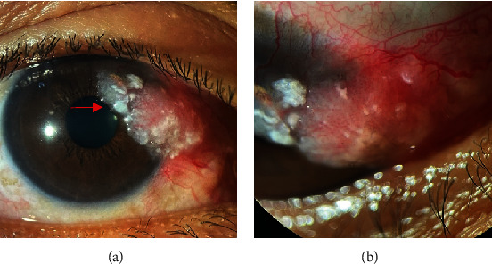
Objective: To describe the morphological and histopathological features of primary conjunctival basal cell carcinoma (BCC) in a young adult Filipino. Introduction: Malignant conjunctival tumors arise from different cells, the most common of which are squamous cell carcinomas (SCCs), (including ocular surface squamous neoplasia [OSSN]), melanomas, and lymphomas. Primary conjunctival BCC is rare and can mimic the clinical features of OSSN. Only seven reported cases were published. Most cases are in the 6th-8th decades of life, and we report the first case in a young adult male. Case Summary: A 37/M, HIV-seronegative, presenting with a 3-year history of enlarging fleshy, pedunculated mass on the right eye measuring 8.5 mm × 8.0 mm at the superonasal limbus encroaching on the cornea, with prominent feeder vessels. Whitish-to-grayish plaques were observed on the surface of the lesions. Wide excision of the mass using the no-touch technique was performed under local anesthesia. Four cycles of mitomycin C (0.02%) were administered as chemoadjuvant therapy. Histopathology showed basaloid cells with peripheral palisading, most consistent with BCC. Immunohistochemistry was positive for Bcl-2 and CD10 markers and negative for epithelial membrane antigen (EMA) and carcinoembryonic antigen (CEA), confirming conjunctival BCC. Eight weeks postoperatively, fibrovascular tissue proliferation was noted at the excision site. Anterior segment-optical coherence tomography (AS-OCT) revealed a thickened hyperreflective band that was continuous with the epithelium, indicating possible recurrence. Resection with rush frozen section revealed fibrotic tissue that was negative for tumor cells. The bare sclera was covered with conjunctival autograft. There was no recurrence of the lesion after 16 months of follow-up. Conclusion: Primary BCC of the conjunctiva is rarely encountered, especially in young individuals, mimicking squamous neoplasia both in morphology and histopathology. Therefore, this should be considered in the differential diagnosis of OSSN. Immunostaining is crucial in differentiating between the two conditions and confirming the diagnosis. In most cases, wide surgical excision is sufficient. In addition, adjuvant therapies may be beneficial in preventing tumor recurrence.

 求助内容:
求助内容: 应助结果提醒方式:


