Pierre Cnockaert , Gregory Reychler , Renaud Menten , Jan Steckel , William Poncin
{"title":"使用主流桌面 3D 打印机制作的小儿上气道模型的几何验证","authors":"Pierre Cnockaert , Gregory Reychler , Renaud Menten , Jan Steckel , William Poncin","doi":"10.1016/j.stlm.2024.100165","DOIUrl":null,"url":null,"abstract":"<div><h3>Background</h3><p>Three-dimensional (3D) printing has become increasingly affordable. Several research projects used 3D printing to create in vitro upper airways model. However, studies using a mainstream desktop 3D printer never performed geometric validation of their model. The aim of this study was to perform geometric validation of a pediatric upper airways model printed with a mainstream desktop 3D printer.</p></div><div><h3>Methods</h3><p>Head computerized tomography (CT) scan of a 10-month-old female underwent segmentation between airways and surrounding anatomical structures. Airways segmentation allowed their measurement for further comparison with printed model. Head segmentation enabled the creation of a 3D printable volume file. To proceed to the geometric validation of the head model, the latter underwent a CT scan. Similar segmentation work was performed on the printed model for comparison. Overlap proportion between the original infant volume and the printed model as well as an average Hausdorff distance were calculated after manual alignment between the original and printed model.</p></div><div><h3>Results</h3><p>Volumes were 12.31 cm<sup>3</sup> and 12.32 cm<sup>3</sup> for the pediatric and the printed model upper airways, respectively (0.08% difference). Dice coefficient of original and printed model was 0.92. The average Hausdorff distance was 0.21 mm.</p></div><div><h3>Conclusion</h3><p>Desktop mainstream 3D printers can generate pediatric upper airway model with a high dimensional accuracy, as evidenced by our comprehensive geometrical validation.</p></div>","PeriodicalId":72210,"journal":{"name":"Annals of 3D printed medicine","volume":"15 ","pages":"Article 100165"},"PeriodicalIF":0.0000,"publicationDate":"2024-07-10","publicationTypes":"Journal Article","fieldsOfStudy":null,"isOpenAccess":false,"openAccessPdf":"https://www.sciencedirect.com/science/article/pii/S2666964124000249/pdfft?md5=8cfe714703dbecce3e19adc0e2c0c990&pid=1-s2.0-S2666964124000249-main.pdf","citationCount":"0","resultStr":"{\"title\":\"Geometric validation of a pediatric upper airways model made using a mainstream desktop 3D printer\",\"authors\":\"Pierre Cnockaert , Gregory Reychler , Renaud Menten , Jan Steckel , William Poncin\",\"doi\":\"10.1016/j.stlm.2024.100165\",\"DOIUrl\":null,\"url\":null,\"abstract\":\"<div><h3>Background</h3><p>Three-dimensional (3D) printing has become increasingly affordable. Several research projects used 3D printing to create in vitro upper airways model. However, studies using a mainstream desktop 3D printer never performed geometric validation of their model. The aim of this study was to perform geometric validation of a pediatric upper airways model printed with a mainstream desktop 3D printer.</p></div><div><h3>Methods</h3><p>Head computerized tomography (CT) scan of a 10-month-old female underwent segmentation between airways and surrounding anatomical structures. Airways segmentation allowed their measurement for further comparison with printed model. Head segmentation enabled the creation of a 3D printable volume file. To proceed to the geometric validation of the head model, the latter underwent a CT scan. Similar segmentation work was performed on the printed model for comparison. Overlap proportion between the original infant volume and the printed model as well as an average Hausdorff distance were calculated after manual alignment between the original and printed model.</p></div><div><h3>Results</h3><p>Volumes were 12.31 cm<sup>3</sup> and 12.32 cm<sup>3</sup> for the pediatric and the printed model upper airways, respectively (0.08% difference). Dice coefficient of original and printed model was 0.92. The average Hausdorff distance was 0.21 mm.</p></div><div><h3>Conclusion</h3><p>Desktop mainstream 3D printers can generate pediatric upper airway model with a high dimensional accuracy, as evidenced by our comprehensive geometrical validation.</p></div>\",\"PeriodicalId\":72210,\"journal\":{\"name\":\"Annals of 3D printed medicine\",\"volume\":\"15 \",\"pages\":\"Article 100165\"},\"PeriodicalIF\":0.0000,\"publicationDate\":\"2024-07-10\",\"publicationTypes\":\"Journal Article\",\"fieldsOfStudy\":null,\"isOpenAccess\":false,\"openAccessPdf\":\"https://www.sciencedirect.com/science/article/pii/S2666964124000249/pdfft?md5=8cfe714703dbecce3e19adc0e2c0c990&pid=1-s2.0-S2666964124000249-main.pdf\",\"citationCount\":\"0\",\"resultStr\":null,\"platform\":\"Semanticscholar\",\"paperid\":null,\"PeriodicalName\":\"Annals of 3D printed medicine\",\"FirstCategoryId\":\"1085\",\"ListUrlMain\":\"https://www.sciencedirect.com/science/article/pii/S2666964124000249\",\"RegionNum\":0,\"RegionCategory\":null,\"ArticlePicture\":[],\"TitleCN\":null,\"AbstractTextCN\":null,\"PMCID\":null,\"EPubDate\":\"\",\"PubModel\":\"\",\"JCR\":\"Q3\",\"JCRName\":\"Medicine\",\"Score\":null,\"Total\":0}","platform":"Semanticscholar","paperid":null,"PeriodicalName":"Annals of 3D printed medicine","FirstCategoryId":"1085","ListUrlMain":"https://www.sciencedirect.com/science/article/pii/S2666964124000249","RegionNum":0,"RegionCategory":null,"ArticlePicture":[],"TitleCN":null,"AbstractTextCN":null,"PMCID":null,"EPubDate":"","PubModel":"","JCR":"Q3","JCRName":"Medicine","Score":null,"Total":0}
引用次数: 0
摘要
背景三维(3D)打印变得越来越经济实惠。一些研究项目使用三维打印技术制作体外上呼吸道模型。然而,使用主流桌面 3D 打印机的研究从未对其模型进行几何验证。本研究的目的是对使用主流桌面 3D 打印机打印的小儿上呼吸道模型进行几何验证。方法对一名 10 个月大女性的头部计算机断层扫描(CT)结果进行气道和周围解剖结构的分割。气道分割后可对其进行测量,以便与打印模型进行进一步比较。对头部进行分割后,就可以创建可打印的三维体积文件。为了对头部模型进行几何验证,后者接受了 CT 扫描。在打印模型上也进行了类似的分割工作,以便进行比较。在对原始模型和打印模型进行手动对齐后,计算了原始婴儿体积和打印模型之间的重叠比例以及平均豪斯多夫距离。结果小儿和打印模型上气道的体积分别为 12.31 立方厘米和 12.32 立方厘米(相差 0.08%)。原始模型和印刷模型的骰子系数为 0.92。结论桌面主流三维打印机可以生成具有高尺寸精度的儿科上气道模型,我们的综合几何验证也证明了这一点。
Geometric validation of a pediatric upper airways model made using a mainstream desktop 3D printer
Background
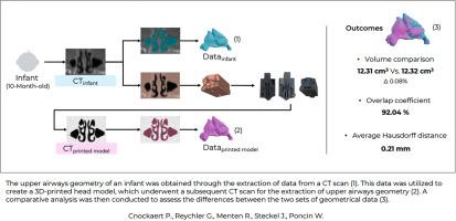
Three-dimensional (3D) printing has become increasingly affordable. Several research projects used 3D printing to create in vitro upper airways model. However, studies using a mainstream desktop 3D printer never performed geometric validation of their model. The aim of this study was to perform geometric validation of a pediatric upper airways model printed with a mainstream desktop 3D printer.
Methods
Head computerized tomography (CT) scan of a 10-month-old female underwent segmentation between airways and surrounding anatomical structures. Airways segmentation allowed their measurement for further comparison with printed model. Head segmentation enabled the creation of a 3D printable volume file. To proceed to the geometric validation of the head model, the latter underwent a CT scan. Similar segmentation work was performed on the printed model for comparison. Overlap proportion between the original infant volume and the printed model as well as an average Hausdorff distance were calculated after manual alignment between the original and printed model.
Results
Volumes were 12.31 cm3 and 12.32 cm3 for the pediatric and the printed model upper airways, respectively (0.08% difference). Dice coefficient of original and printed model was 0.92. The average Hausdorff distance was 0.21 mm.
Conclusion
Desktop mainstream 3D printers can generate pediatric upper airway model with a high dimensional accuracy, as evidenced by our comprehensive geometrical validation.

 求助内容:
求助内容: 应助结果提醒方式:


