{"title":"胎儿神经超声显示孤立性脑室肥大的脉络丛生长是否发生改变?","authors":"Wei-Xi Hu, Xin Zhan, Dan Lu, Zhi-Qiang Li","doi":"10.1007/s00330-024-10966-3","DOIUrl":null,"url":null,"abstract":"<p><strong>Objectives: </strong>Reveal developmental alterations in choroid plexus volume (CPV) among fetuses with isolated ventriculomegaly (VM) through neuro-ultrasound.</p><p><strong>Methods: </strong>This prospective study aimed to assess the development of fetal CPV in normal fetuses and those with isolated VM through neuro-ultrasound. The fetuses of isolated VM were categorized into mild, moderate, and severe groups, and subsequently, the lateral ventricle evolution was monitored. The developmental alterations in CPV among fetuses with isolated VM were determined by comparing the CPV z-scores with those of normal fetuses. Receiver operating characteristics curve analysis was used to assess the predictive value of altered CPV in lateral ventricle evolution.</p><p><strong>Results: </strong>A total of 218 normal fetuses and 114 isolated VM fetuses from 22 weeks to 35 weeks of gestation were included. The CPV decreased as the isolated VM was getting worse. Both fetuses with isolated moderate ventriculomegaly and those with isolated severe ventriculomegaly exhibited reduced CPV compared to normal fetuses. The CPV in fetuses with isolated mild ventriculomegaly (IMVM) varied, with some showing a larger CPV compared to normal fetuses, while others exhibited a smaller CPV. The larger CPV in cases of IMVM may serve as a predictive factor for either regression or stability of the lateral ventricle, while reduced CPV in cases of isolated VM may indicate worsening of the lateral ventricle.</p><p><strong>Conclusion: </strong>The growth volume of fetal CP exhibited alterations in fetuses with isolated VM, and these changes were found to be correlated with the evolution of the lateral ventricle.</p><p><strong>Clinical relevance statement: </strong>Neuro-ultrasound revealed varying degrees of alterations in the volume development of the choroid plexus within the fetus with isolated VM. The findings can help predict lateral ventricle prognosis, greatly contributing to prenatal diagnosis strategies for fetuses with isolated VM.</p><p><strong>Key points: </strong>The volume of choroid plexus growth is altered in fetuses with isolated VM. The altered CPV in isolated VM was associated with lateral ventricle evolution. The findings are useful for prenatal counseling and managing fetuses with isolated VM.</p>","PeriodicalId":12076,"journal":{"name":"European Radiology","volume":" ","pages":"463-473"},"PeriodicalIF":4.7000,"publicationDate":"2025-01-01","publicationTypes":"Journal Article","fieldsOfStudy":null,"isOpenAccess":false,"openAccessPdf":"https://www.ncbi.nlm.nih.gov/pmc/articles/PMC11632053/pdf/","citationCount":"0","resultStr":"{\"title\":\"Is choroid plexus growth altered in isolated ventriculomegaly on fetal neuro-ultrasound?\",\"authors\":\"Wei-Xi Hu, Xin Zhan, Dan Lu, Zhi-Qiang Li\",\"doi\":\"10.1007/s00330-024-10966-3\",\"DOIUrl\":null,\"url\":null,\"abstract\":\"<p><strong>Objectives: </strong>Reveal developmental alterations in choroid plexus volume (CPV) among fetuses with isolated ventriculomegaly (VM) through neuro-ultrasound.</p><p><strong>Methods: </strong>This prospective study aimed to assess the development of fetal CPV in normal fetuses and those with isolated VM through neuro-ultrasound. The fetuses of isolated VM were categorized into mild, moderate, and severe groups, and subsequently, the lateral ventricle evolution was monitored. The developmental alterations in CPV among fetuses with isolated VM were determined by comparing the CPV z-scores with those of normal fetuses. Receiver operating characteristics curve analysis was used to assess the predictive value of altered CPV in lateral ventricle evolution.</p><p><strong>Results: </strong>A total of 218 normal fetuses and 114 isolated VM fetuses from 22 weeks to 35 weeks of gestation were included. The CPV decreased as the isolated VM was getting worse. Both fetuses with isolated moderate ventriculomegaly and those with isolated severe ventriculomegaly exhibited reduced CPV compared to normal fetuses. The CPV in fetuses with isolated mild ventriculomegaly (IMVM) varied, with some showing a larger CPV compared to normal fetuses, while others exhibited a smaller CPV. The larger CPV in cases of IMVM may serve as a predictive factor for either regression or stability of the lateral ventricle, while reduced CPV in cases of isolated VM may indicate worsening of the lateral ventricle.</p><p><strong>Conclusion: </strong>The growth volume of fetal CP exhibited alterations in fetuses with isolated VM, and these changes were found to be correlated with the evolution of the lateral ventricle.</p><p><strong>Clinical relevance statement: </strong>Neuro-ultrasound revealed varying degrees of alterations in the volume development of the choroid plexus within the fetus with isolated VM. The findings can help predict lateral ventricle prognosis, greatly contributing to prenatal diagnosis strategies for fetuses with isolated VM.</p><p><strong>Key points: </strong>The volume of choroid plexus growth is altered in fetuses with isolated VM. The altered CPV in isolated VM was associated with lateral ventricle evolution. The findings are useful for prenatal counseling and managing fetuses with isolated VM.</p>\",\"PeriodicalId\":12076,\"journal\":{\"name\":\"European Radiology\",\"volume\":\" \",\"pages\":\"463-473\"},\"PeriodicalIF\":4.7000,\"publicationDate\":\"2025-01-01\",\"publicationTypes\":\"Journal Article\",\"fieldsOfStudy\":null,\"isOpenAccess\":false,\"openAccessPdf\":\"https://www.ncbi.nlm.nih.gov/pmc/articles/PMC11632053/pdf/\",\"citationCount\":\"0\",\"resultStr\":null,\"platform\":\"Semanticscholar\",\"paperid\":null,\"PeriodicalName\":\"European Radiology\",\"FirstCategoryId\":\"3\",\"ListUrlMain\":\"https://doi.org/10.1007/s00330-024-10966-3\",\"RegionNum\":2,\"RegionCategory\":\"医学\",\"ArticlePicture\":[],\"TitleCN\":null,\"AbstractTextCN\":null,\"PMCID\":null,\"EPubDate\":\"2024/7/17 0:00:00\",\"PubModel\":\"Epub\",\"JCR\":\"Q1\",\"JCRName\":\"RADIOLOGY, NUCLEAR MEDICINE & MEDICAL IMAGING\",\"Score\":null,\"Total\":0}","platform":"Semanticscholar","paperid":null,"PeriodicalName":"European Radiology","FirstCategoryId":"3","ListUrlMain":"https://doi.org/10.1007/s00330-024-10966-3","RegionNum":2,"RegionCategory":"医学","ArticlePicture":[],"TitleCN":null,"AbstractTextCN":null,"PMCID":null,"EPubDate":"2024/7/17 0:00:00","PubModel":"Epub","JCR":"Q1","JCRName":"RADIOLOGY, NUCLEAR MEDICINE & MEDICAL IMAGING","Score":null,"Total":0}
引用次数: 0
摘要
目的:通过神经超声揭示孤立性脑室肥大(VM)胎儿脉络丛体积(CPV)的发育改变:通过神经超声揭示孤立性脑室肥大(VM)胎儿脉络丛体积(CPV)的发育改变:这项前瞻性研究旨在通过神经超声评估正常胎儿和孤立性脑室肥大胎儿的脉络丛体积发育情况。将孤立性 VM 胎儿分为轻度、中度和重度组,随后监测侧脑室的演变。通过比较孤立性 VM 胎儿与正常胎儿的 CPV z 评分,确定 CPV 的发育改变。采用受试者操作特征曲线分析评估 CPV 改变对侧脑室演变的预测价值:结果:共纳入了妊娠 22 周至 35 周的 218 个正常胎儿和 114 个孤立 VM 胎儿。CPV随着孤立性脑室病变的加重而下降。与正常胎儿相比,孤立性中度脑室肥大和孤立性重度脑室肥大胎儿的 CPV 均有所下降。与正常胎儿相比,孤立性轻度脑室肥大(IMVM)胎儿的 CPV 各不相同,有些胎儿的 CPV 较大,有些则较小。IMVM病例中CPV较大可能是侧脑室退变或稳定的预测因素,而孤立性轻度脑室肥大病例中CPV减小可能表明侧脑室恶化:结论:在孤立性 VM 胎儿中,胎儿 CP 的生长体积发生了变化,这些变化与侧脑室的演变相关:神经超声揭示了孤立性VM胎儿脉络丛体积发育的不同程度改变。研究结果有助于预测侧脑室的预后,对孤立性 VM 胎儿的产前诊断策略大有裨益:要点:孤立性VM胎儿的脉络丛生长体积发生改变。要点:孤立性 VM 胎儿的脉络丛生长体积发生改变,孤立性 VM 胎儿脉络丛生长体积的改变与侧脑室的演变有关。这些发现有助于产前咨询和管理孤立性血管瘤胎儿。
Is choroid plexus growth altered in isolated ventriculomegaly on fetal neuro-ultrasound?
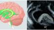
Objectives: Reveal developmental alterations in choroid plexus volume (CPV) among fetuses with isolated ventriculomegaly (VM) through neuro-ultrasound.
Methods: This prospective study aimed to assess the development of fetal CPV in normal fetuses and those with isolated VM through neuro-ultrasound. The fetuses of isolated VM were categorized into mild, moderate, and severe groups, and subsequently, the lateral ventricle evolution was monitored. The developmental alterations in CPV among fetuses with isolated VM were determined by comparing the CPV z-scores with those of normal fetuses. Receiver operating characteristics curve analysis was used to assess the predictive value of altered CPV in lateral ventricle evolution.
Results: A total of 218 normal fetuses and 114 isolated VM fetuses from 22 weeks to 35 weeks of gestation were included. The CPV decreased as the isolated VM was getting worse. Both fetuses with isolated moderate ventriculomegaly and those with isolated severe ventriculomegaly exhibited reduced CPV compared to normal fetuses. The CPV in fetuses with isolated mild ventriculomegaly (IMVM) varied, with some showing a larger CPV compared to normal fetuses, while others exhibited a smaller CPV. The larger CPV in cases of IMVM may serve as a predictive factor for either regression or stability of the lateral ventricle, while reduced CPV in cases of isolated VM may indicate worsening of the lateral ventricle.
Conclusion: The growth volume of fetal CP exhibited alterations in fetuses with isolated VM, and these changes were found to be correlated with the evolution of the lateral ventricle.
Clinical relevance statement: Neuro-ultrasound revealed varying degrees of alterations in the volume development of the choroid plexus within the fetus with isolated VM. The findings can help predict lateral ventricle prognosis, greatly contributing to prenatal diagnosis strategies for fetuses with isolated VM.
Key points: The volume of choroid plexus growth is altered in fetuses with isolated VM. The altered CPV in isolated VM was associated with lateral ventricle evolution. The findings are useful for prenatal counseling and managing fetuses with isolated VM.
期刊介绍:
European Radiology (ER) continuously updates scientific knowledge in radiology by publication of strong original articles and state-of-the-art reviews written by leading radiologists. A well balanced combination of review articles, original papers, short communications from European radiological congresses and information on society matters makes ER an indispensable source for current information in this field.
This is the Journal of the European Society of Radiology, and the official journal of a number of societies.
From 2004-2008 supplements to European Radiology were published under its companion, European Radiology Supplements, ISSN 1613-3749.

 求助内容:
求助内容: 应助结果提醒方式:


