Michael A Krause, Marta Grannonico, Brooke P Tyler, David A Miller, Weijia Fan, Mingna Liu, Roman V Kuranov, Hao F Zhang, Xiaorong Liu, Peter A Netland
{"title":"通过可见光光学相干断层扫描的新应用观察眼窝中央的超反射点。","authors":"Michael A Krause, Marta Grannonico, Brooke P Tyler, David A Miller, Weijia Fan, Mingna Liu, Roman V Kuranov, Hao F Zhang, Xiaorong Liu, Peter A Netland","doi":"10.1155/2024/5823455","DOIUrl":null,"url":null,"abstract":"<p><p>Visible-light optical coherence tomography (vis-OCT) is a novel noninvasive retinal imaging system that offers improved resolution compared to conventional near-infrared (NIR) OCT systems. Here, we utilized vis-OCT to produce fibergrams (vis-OCTF) for the first time in human patients, enabling <i>en face</i> visualization and precise quantification of hyperreflective dots in the central fovea in two patients. We also directly compare the imaging qualities of conventional vis-OCT and NIR-OCT. Vis-OCT generated a 3 × 3 mm<sup>2</sup> <i>en face</i> image with an impressive axial resolution of 1.3 <i>μ</i>m, whereas NIR-OCT produced an <i>en face</i> image with a larger field of view (FOV) (9 × 9 mm<sup>2</sup>) but a lower resolution of 7.0 <i>μ</i>m. Moreover, vis-OCTF unveiled clear images of hyperreflective dots in the fovea of both patients, which were not discernible in the NIR-OCT <i>en face</i> images. Foveal dots have often been linked to several age-related and pathological conditions. The high-resolution images generated by vis-OCTF enable more precise characterization of changes in retinal sublayers within the central fovea.</p>","PeriodicalId":9603,"journal":{"name":"Case Reports in Ophthalmological Medicine","volume":"2024 ","pages":"5823455"},"PeriodicalIF":0.4000,"publicationDate":"2024-07-09","publicationTypes":"Journal Article","fieldsOfStudy":null,"isOpenAccess":false,"openAccessPdf":"https://www.ncbi.nlm.nih.gov/pmc/articles/PMC11251792/pdf/","citationCount":"0","resultStr":"{\"title\":\"Hyperreflective Dots in Central Fovea Visualized by a Novel Application of Visible-Light Optical Coherence Tomography.\",\"authors\":\"Michael A Krause, Marta Grannonico, Brooke P Tyler, David A Miller, Weijia Fan, Mingna Liu, Roman V Kuranov, Hao F Zhang, Xiaorong Liu, Peter A Netland\",\"doi\":\"10.1155/2024/5823455\",\"DOIUrl\":null,\"url\":null,\"abstract\":\"<p><p>Visible-light optical coherence tomography (vis-OCT) is a novel noninvasive retinal imaging system that offers improved resolution compared to conventional near-infrared (NIR) OCT systems. Here, we utilized vis-OCT to produce fibergrams (vis-OCTF) for the first time in human patients, enabling <i>en face</i> visualization and precise quantification of hyperreflective dots in the central fovea in two patients. We also directly compare the imaging qualities of conventional vis-OCT and NIR-OCT. Vis-OCT generated a 3 × 3 mm<sup>2</sup> <i>en face</i> image with an impressive axial resolution of 1.3 <i>μ</i>m, whereas NIR-OCT produced an <i>en face</i> image with a larger field of view (FOV) (9 × 9 mm<sup>2</sup>) but a lower resolution of 7.0 <i>μ</i>m. Moreover, vis-OCTF unveiled clear images of hyperreflective dots in the fovea of both patients, which were not discernible in the NIR-OCT <i>en face</i> images. Foveal dots have often been linked to several age-related and pathological conditions. The high-resolution images generated by vis-OCTF enable more precise characterization of changes in retinal sublayers within the central fovea.</p>\",\"PeriodicalId\":9603,\"journal\":{\"name\":\"Case Reports in Ophthalmological Medicine\",\"volume\":\"2024 \",\"pages\":\"5823455\"},\"PeriodicalIF\":0.4000,\"publicationDate\":\"2024-07-09\",\"publicationTypes\":\"Journal Article\",\"fieldsOfStudy\":null,\"isOpenAccess\":false,\"openAccessPdf\":\"https://www.ncbi.nlm.nih.gov/pmc/articles/PMC11251792/pdf/\",\"citationCount\":\"0\",\"resultStr\":null,\"platform\":\"Semanticscholar\",\"paperid\":null,\"PeriodicalName\":\"Case Reports in Ophthalmological Medicine\",\"FirstCategoryId\":\"1085\",\"ListUrlMain\":\"https://doi.org/10.1155/2024/5823455\",\"RegionNum\":0,\"RegionCategory\":null,\"ArticlePicture\":[],\"TitleCN\":null,\"AbstractTextCN\":null,\"PMCID\":null,\"EPubDate\":\"2024/1/1 0:00:00\",\"PubModel\":\"eCollection\",\"JCR\":\"Q4\",\"JCRName\":\"OPHTHALMOLOGY\",\"Score\":null,\"Total\":0}","platform":"Semanticscholar","paperid":null,"PeriodicalName":"Case Reports in Ophthalmological Medicine","FirstCategoryId":"1085","ListUrlMain":"https://doi.org/10.1155/2024/5823455","RegionNum":0,"RegionCategory":null,"ArticlePicture":[],"TitleCN":null,"AbstractTextCN":null,"PMCID":null,"EPubDate":"2024/1/1 0:00:00","PubModel":"eCollection","JCR":"Q4","JCRName":"OPHTHALMOLOGY","Score":null,"Total":0}
Hyperreflective Dots in Central Fovea Visualized by a Novel Application of Visible-Light Optical Coherence Tomography.
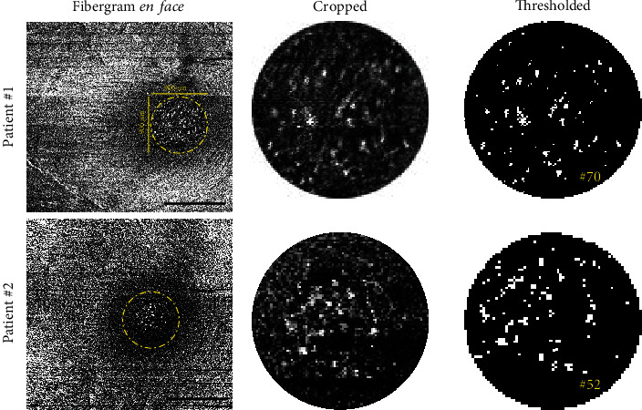
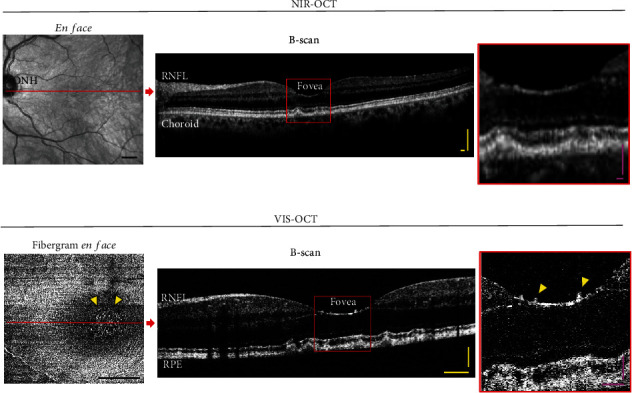
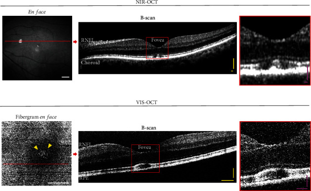
Visible-light optical coherence tomography (vis-OCT) is a novel noninvasive retinal imaging system that offers improved resolution compared to conventional near-infrared (NIR) OCT systems. Here, we utilized vis-OCT to produce fibergrams (vis-OCTF) for the first time in human patients, enabling en face visualization and precise quantification of hyperreflective dots in the central fovea in two patients. We also directly compare the imaging qualities of conventional vis-OCT and NIR-OCT. Vis-OCT generated a 3 × 3 mm2en face image with an impressive axial resolution of 1.3 μm, whereas NIR-OCT produced an en face image with a larger field of view (FOV) (9 × 9 mm2) but a lower resolution of 7.0 μm. Moreover, vis-OCTF unveiled clear images of hyperreflective dots in the fovea of both patients, which were not discernible in the NIR-OCT en face images. Foveal dots have often been linked to several age-related and pathological conditions. The high-resolution images generated by vis-OCTF enable more precise characterization of changes in retinal sublayers within the central fovea.

 求助内容:
求助内容: 应助结果提醒方式:


