Changpeng Chai, Huan Tang, Xin Miao, Yuanhui Su, Lu Li, Cheng Yu, Jianfeng Yi, Zhenzhen Ye, Long Miao, Bo Zhang, Zhengfeng Wang, Wei Luo, Jinjing Hu, Hui Zhang, Wence Zhou, Hao Xu
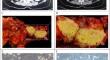
{"title":"PDAC-X3 细胞系的建立和特征描述:一种新型中国来源的胰腺导管腺癌细胞系。","authors":"Changpeng Chai, Huan Tang, Xin Miao, Yuanhui Su, Lu Li, Cheng Yu, Jianfeng Yi, Zhenzhen Ye, Long Miao, Bo Zhang, Zhengfeng Wang, Wei Luo, Jinjing Hu, Hui Zhang, Wence Zhou, Hao Xu","doi":"10.1007/s13577-024-01100-y","DOIUrl":null,"url":null,"abstract":"<p><p>In this study, a novel pancreatic cancer cell line, termed pancreatic ductal adenocarcinoma (PDAC)-X3 cell line, was successfully derived from the primary tumor. Comprehensive analyses of its malignant phenotype, molecular properties, specific biomarkers, and histological features confirmed that PDAC-X3 cells serve as a valuable model for investigating the underlying mechanisms driving pancreatic carcinogenesis and advancing potential therapeutic strategies. The newly established cell line was continuously cultured for over 12 months and was stably passaged through more than 50 generations. Morphologically, PDAC-X3 cells displayed characteristics typical of epithelial tumors. The population doubling time for PDAC-X3 cells was determined to be 50 h. Karyotype analysis revealed that 75% of PDAC-X3 cells presented as hypotriploid, while 25% were sub-tetraploid, with representative karyotypes being 53 and XY der (1) inv (9) der (22). In suspension culture, PDAC-X3 cells efficiently formed organoids. Upon inoculation into BALB/C nude mice, these cells initiated the development of xenograft tumors, achieving a tumor formation rate of 33%. Morphologically, these xenografted tumors closely resembled the primary tumor. Drug sensitivity assays indicated that PDAC-X3 cells exhibited resistance to oxaliplatin but demonstrated sensitivity to 5-Fluorouracil (5-FU), gemcitabine, and paclitaxel. Immunohistochemical analysis revealed that CK7, CK19, E-cadherin, Vimentin, CA19-9 were positively expressed in PDAC-X3 cells. Meanwhile, the expression rate for Ki-67 was 30%, and that for CEA was not detected. Our findings underscore that PDAC-X3 represents a novel pancreatic cancer cell line, positioning it as a valuable model for basic research and the advancement of therapeutic strategies against pancreatic cancer.</p>","PeriodicalId":49194,"journal":{"name":"Human Cell","volume":" ","pages":"1578-1592"},"PeriodicalIF":3.4000,"publicationDate":"2024-09-01","publicationTypes":"Journal Article","fieldsOfStudy":null,"isOpenAccess":false,"openAccessPdf":"","citationCount":"0","resultStr":"{\"title\":\"Establishment and characterization of the PDAC-X3 cell line: a novel Chinese-origin pancreatic ductal adenocarcinoma cell line.\",\"authors\":\"Changpeng Chai, Huan Tang, Xin Miao, Yuanhui Su, Lu Li, Cheng Yu, Jianfeng Yi, Zhenzhen Ye, Long Miao, Bo Zhang, Zhengfeng Wang, Wei Luo, Jinjing Hu, Hui Zhang, Wence Zhou, Hao Xu\",\"doi\":\"10.1007/s13577-024-01100-y\",\"DOIUrl\":null,\"url\":null,\"abstract\":\"<p><p>In this study, a novel pancreatic cancer cell line, termed pancreatic ductal adenocarcinoma (PDAC)-X3 cell line, was successfully derived from the primary tumor. Comprehensive analyses of its malignant phenotype, molecular properties, specific biomarkers, and histological features confirmed that PDAC-X3 cells serve as a valuable model for investigating the underlying mechanisms driving pancreatic carcinogenesis and advancing potential therapeutic strategies. The newly established cell line was continuously cultured for over 12 months and was stably passaged through more than 50 generations. Morphologically, PDAC-X3 cells displayed characteristics typical of epithelial tumors. The population doubling time for PDAC-X3 cells was determined to be 50 h. Karyotype analysis revealed that 75% of PDAC-X3 cells presented as hypotriploid, while 25% were sub-tetraploid, with representative karyotypes being 53 and XY der (1) inv (9) der (22). In suspension culture, PDAC-X3 cells efficiently formed organoids. Upon inoculation into BALB/C nude mice, these cells initiated the development of xenograft tumors, achieving a tumor formation rate of 33%. Morphologically, these xenografted tumors closely resembled the primary tumor. Drug sensitivity assays indicated that PDAC-X3 cells exhibited resistance to oxaliplatin but demonstrated sensitivity to 5-Fluorouracil (5-FU), gemcitabine, and paclitaxel. Immunohistochemical analysis revealed that CK7, CK19, E-cadherin, Vimentin, CA19-9 were positively expressed in PDAC-X3 cells. Meanwhile, the expression rate for Ki-67 was 30%, and that for CEA was not detected. Our findings underscore that PDAC-X3 represents a novel pancreatic cancer cell line, positioning it as a valuable model for basic research and the advancement of therapeutic strategies against pancreatic cancer.</p>\",\"PeriodicalId\":49194,\"journal\":{\"name\":\"Human Cell\",\"volume\":\" \",\"pages\":\"1578-1592\"},\"PeriodicalIF\":3.4000,\"publicationDate\":\"2024-09-01\",\"publicationTypes\":\"Journal Article\",\"fieldsOfStudy\":null,\"isOpenAccess\":false,\"openAccessPdf\":\"\",\"citationCount\":\"0\",\"resultStr\":null,\"platform\":\"Semanticscholar\",\"paperid\":null,\"PeriodicalName\":\"Human Cell\",\"FirstCategoryId\":\"99\",\"ListUrlMain\":\"https://doi.org/10.1007/s13577-024-01100-y\",\"RegionNum\":3,\"RegionCategory\":\"生物学\",\"ArticlePicture\":[],\"TitleCN\":null,\"AbstractTextCN\":null,\"PMCID\":null,\"EPubDate\":\"2024/7/16 0:00:00\",\"PubModel\":\"Epub\",\"JCR\":\"Q3\",\"JCRName\":\"CELL BIOLOGY\",\"Score\":null,\"Total\":0}","platform":"Semanticscholar","paperid":null,"PeriodicalName":"Human Cell","FirstCategoryId":"99","ListUrlMain":"https://doi.org/10.1007/s13577-024-01100-y","RegionNum":3,"RegionCategory":"生物学","ArticlePicture":[],"TitleCN":null,"AbstractTextCN":null,"PMCID":null,"EPubDate":"2024/7/16 0:00:00","PubModel":"Epub","JCR":"Q3","JCRName":"CELL BIOLOGY","Score":null,"Total":0}
Establishment and characterization of the PDAC-X3 cell line: a novel Chinese-origin pancreatic ductal adenocarcinoma cell line.
In this study, a novel pancreatic cancer cell line, termed pancreatic ductal adenocarcinoma (PDAC)-X3 cell line, was successfully derived from the primary tumor. Comprehensive analyses of its malignant phenotype, molecular properties, specific biomarkers, and histological features confirmed that PDAC-X3 cells serve as a valuable model for investigating the underlying mechanisms driving pancreatic carcinogenesis and advancing potential therapeutic strategies. The newly established cell line was continuously cultured for over 12 months and was stably passaged through more than 50 generations. Morphologically, PDAC-X3 cells displayed characteristics typical of epithelial tumors. The population doubling time for PDAC-X3 cells was determined to be 50 h. Karyotype analysis revealed that 75% of PDAC-X3 cells presented as hypotriploid, while 25% were sub-tetraploid, with representative karyotypes being 53 and XY der (1) inv (9) der (22). In suspension culture, PDAC-X3 cells efficiently formed organoids. Upon inoculation into BALB/C nude mice, these cells initiated the development of xenograft tumors, achieving a tumor formation rate of 33%. Morphologically, these xenografted tumors closely resembled the primary tumor. Drug sensitivity assays indicated that PDAC-X3 cells exhibited resistance to oxaliplatin but demonstrated sensitivity to 5-Fluorouracil (5-FU), gemcitabine, and paclitaxel. Immunohistochemical analysis revealed that CK7, CK19, E-cadherin, Vimentin, CA19-9 were positively expressed in PDAC-X3 cells. Meanwhile, the expression rate for Ki-67 was 30%, and that for CEA was not detected. Our findings underscore that PDAC-X3 represents a novel pancreatic cancer cell line, positioning it as a valuable model for basic research and the advancement of therapeutic strategies against pancreatic cancer.
期刊介绍:
Human Cell is the official English-language journal of the Japan Human Cell Society. The journal serves as a forum for international research on all aspects of the human cell, encompassing not only cell biology but also pathology, cytology, and oncology, including clinical oncology. Embryonic stem cells derived from animals, regenerative medicine using animal cells, and experimental animal models with implications for human diseases are covered as well.
Submissions in any of the following categories will be considered: Research Articles, Cell Lines, Rapid Communications, Reviews, and Letters to the Editor. A brief clinical case report focusing on cellular responses to pathological insults in human studies may also be submitted as a Letter to the Editor in a concise and short format.
Not only basic scientists but also gynecologists, oncologists, and other clinical scientists are welcome to submit work expressing new ideas or research using human cells.
 求助内容:
求助内容: 应助结果提醒方式:


