{"title":"缺血再灌注损伤中衰老对胆囊周围腺体的影响","authors":"Kaoru Katano, Shinichi Nakanuma, Tomokazu Tokoro, Ryohei Takei, Satoshi Takada, Mitsuyoshi Okazaki, Kaichiro Kato, Isamu Makino, Kenichi Harada, Shintaro Yagi","doi":"10.1002/jhbp.12047","DOIUrl":null,"url":null,"abstract":"<div>\n \n \n <section>\n \n <h3> Background</h3>\n \n <p>The detailed mechanisms underlying the development of ischemia-type biliary lesions (ITBLs) in aged donor grafts remain unclear. In the present study we aimed to investigate the impact of aging on the response of the peribiliary gland (PBG) to ischemia–reperfusion injury (IRI) and its temporal changes.</p>\n </section>\n \n <section>\n \n <h3> Methods</h3>\n \n <p>Experiments were performed using a 90-min partial warm liver ischemia model in male Wistar rats of two age groups: young (7–8 weeks old) and old (52–60 weeks old). Liver tissues were obtained 24, 72, and 168 h after IRI. Histopathological and immunohistochemical assessments of the perihilar bile duct (PHBD), including the PBG, distal to the clip-clamped site were performed.</p>\n </section>\n \n <section>\n \n <h3> Results</h3>\n \n <p>Young rats showed little change in the bile duct tissues after IRI. However, old rats showed an increased PBG volume in the PHBD and marked PBG cell proliferation 24 h after IRI. Bile duct wall thickening with narrowing of the lumen peaked 72 h after IRI. Mucus production and oxidative stress in the PBG were significantly higher in old than in young rats after IRI. These findings showed a trend toward improvement 168 h after IRI.</p>\n </section>\n \n <section>\n \n <h3> Conclusion</h3>\n \n <p>Age-dependent differences in the response of the PBG to IRI may be related to differences in ITBL frequency.</p>\n </section>\n </div>","PeriodicalId":16056,"journal":{"name":"Journal of Hepato‐Biliary‐Pancreatic Sciences","volume":"31 10","pages":"705-715"},"PeriodicalIF":3.2000,"publicationDate":"2024-07-16","publicationTypes":"Journal Article","fieldsOfStudy":null,"isOpenAccess":false,"openAccessPdf":"","citationCount":"0","resultStr":"{\"title\":\"Impact of aging on peribiliary glands in ischemia–reperfusion injury\",\"authors\":\"Kaoru Katano, Shinichi Nakanuma, Tomokazu Tokoro, Ryohei Takei, Satoshi Takada, Mitsuyoshi Okazaki, Kaichiro Kato, Isamu Makino, Kenichi Harada, Shintaro Yagi\",\"doi\":\"10.1002/jhbp.12047\",\"DOIUrl\":null,\"url\":null,\"abstract\":\"<div>\\n \\n \\n <section>\\n \\n <h3> Background</h3>\\n \\n <p>The detailed mechanisms underlying the development of ischemia-type biliary lesions (ITBLs) in aged donor grafts remain unclear. In the present study we aimed to investigate the impact of aging on the response of the peribiliary gland (PBG) to ischemia–reperfusion injury (IRI) and its temporal changes.</p>\\n </section>\\n \\n <section>\\n \\n <h3> Methods</h3>\\n \\n <p>Experiments were performed using a 90-min partial warm liver ischemia model in male Wistar rats of two age groups: young (7–8 weeks old) and old (52–60 weeks old). Liver tissues were obtained 24, 72, and 168 h after IRI. Histopathological and immunohistochemical assessments of the perihilar bile duct (PHBD), including the PBG, distal to the clip-clamped site were performed.</p>\\n </section>\\n \\n <section>\\n \\n <h3> Results</h3>\\n \\n <p>Young rats showed little change in the bile duct tissues after IRI. However, old rats showed an increased PBG volume in the PHBD and marked PBG cell proliferation 24 h after IRI. Bile duct wall thickening with narrowing of the lumen peaked 72 h after IRI. Mucus production and oxidative stress in the PBG were significantly higher in old than in young rats after IRI. These findings showed a trend toward improvement 168 h after IRI.</p>\\n </section>\\n \\n <section>\\n \\n <h3> Conclusion</h3>\\n \\n <p>Age-dependent differences in the response of the PBG to IRI may be related to differences in ITBL frequency.</p>\\n </section>\\n </div>\",\"PeriodicalId\":16056,\"journal\":{\"name\":\"Journal of Hepato‐Biliary‐Pancreatic Sciences\",\"volume\":\"31 10\",\"pages\":\"705-715\"},\"PeriodicalIF\":3.2000,\"publicationDate\":\"2024-07-16\",\"publicationTypes\":\"Journal Article\",\"fieldsOfStudy\":null,\"isOpenAccess\":false,\"openAccessPdf\":\"\",\"citationCount\":\"0\",\"resultStr\":null,\"platform\":\"Semanticscholar\",\"paperid\":null,\"PeriodicalName\":\"Journal of Hepato‐Biliary‐Pancreatic Sciences\",\"FirstCategoryId\":\"3\",\"ListUrlMain\":\"https://onlinelibrary.wiley.com/doi/10.1002/jhbp.12047\",\"RegionNum\":3,\"RegionCategory\":\"医学\",\"ArticlePicture\":[],\"TitleCN\":null,\"AbstractTextCN\":null,\"PMCID\":null,\"EPubDate\":\"\",\"PubModel\":\"\",\"JCR\":\"Q2\",\"JCRName\":\"GASTROENTEROLOGY & HEPATOLOGY\",\"Score\":null,\"Total\":0}","platform":"Semanticscholar","paperid":null,"PeriodicalName":"Journal of Hepato‐Biliary‐Pancreatic Sciences","FirstCategoryId":"3","ListUrlMain":"https://onlinelibrary.wiley.com/doi/10.1002/jhbp.12047","RegionNum":3,"RegionCategory":"医学","ArticlePicture":[],"TitleCN":null,"AbstractTextCN":null,"PMCID":null,"EPubDate":"","PubModel":"","JCR":"Q2","JCRName":"GASTROENTEROLOGY & HEPATOLOGY","Score":null,"Total":0}
引用次数: 0
摘要
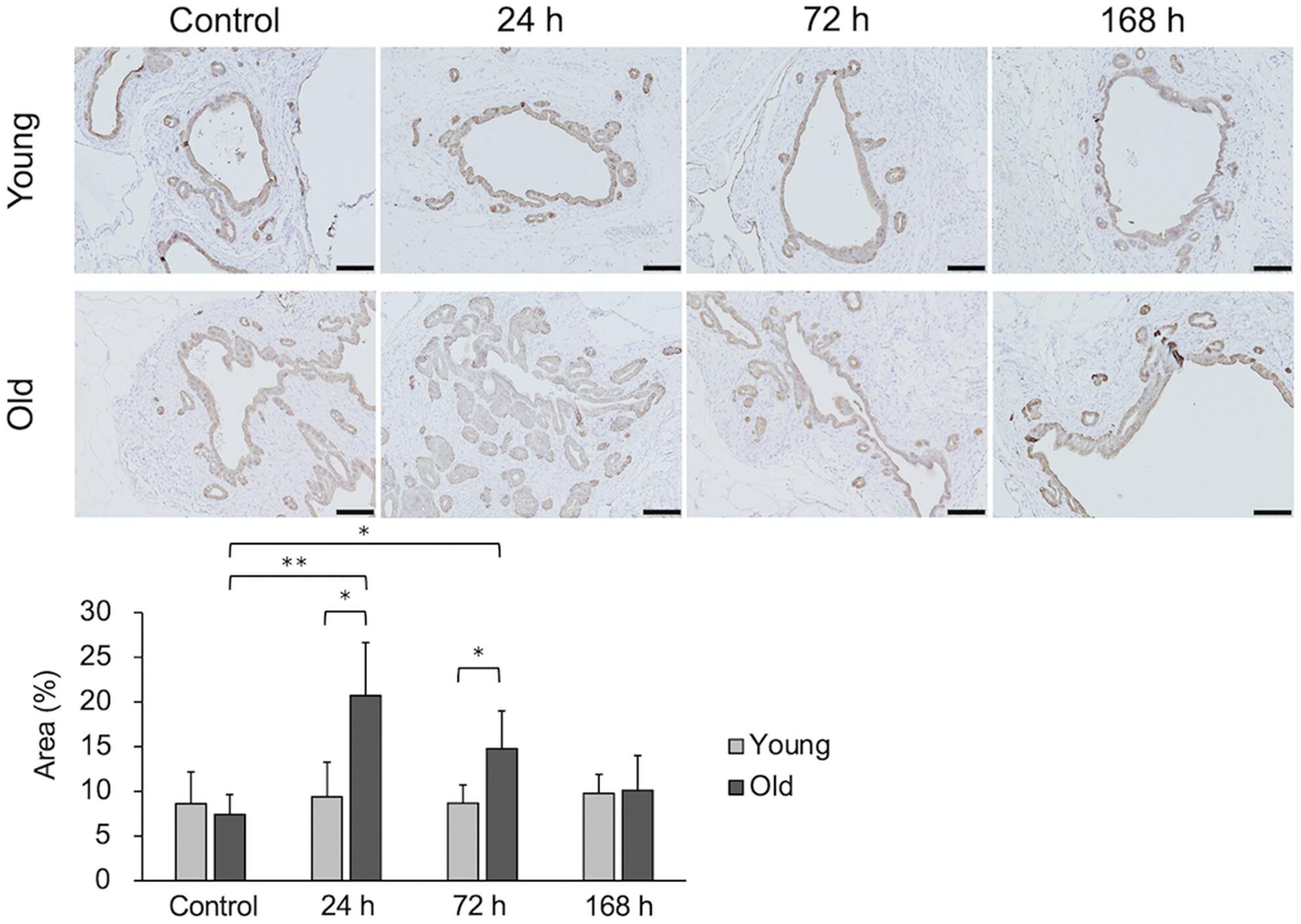
背景:高龄供体移植物缺血型胆道病变(ITBLs)发生的详细机制仍不清楚。本研究旨在探讨衰老对胆管周围腺体(PBG)缺血再灌注损伤(IRI)反应的影响及其时间变化:方法:实验采用 90 分钟部分温热肝缺血模型,分别在年轻(7-8 周龄)和年老(52-60 周龄)两组雄性 Wistar 大鼠中进行。分别在 IRI 后 24、72 和 168 小时采集肝组织。对夹闭部位远端包括 PBG 在内的肝周胆管 (PHBD) 进行组织病理学和免疫组化评估:结果:IRI后,年轻大鼠的胆管组织变化不大。结果:IRI 后,年轻大鼠的胆管组织变化不大,但老年大鼠 PHBD 的 PBG 体积增大,且在 IRI 24 小时后 PBG 细胞明显增殖。胆管壁增厚和管腔变窄在 IRI 72 小时后达到高峰。IRI 后,老龄大鼠 PBG 的粘液分泌和氧化应激明显高于年轻大鼠。这些结果表明,在 IRI 168 小时后,情况有改善的趋势:结论:PBG 对 IRI 反应的年龄依赖性差异可能与 ITBL 频率的差异有关。
Impact of aging on peribiliary glands in ischemia–reperfusion injury
Background
The detailed mechanisms underlying the development of ischemia-type biliary lesions (ITBLs) in aged donor grafts remain unclear. In the present study we aimed to investigate the impact of aging on the response of the peribiliary gland (PBG) to ischemia–reperfusion injury (IRI) and its temporal changes.
Methods
Experiments were performed using a 90-min partial warm liver ischemia model in male Wistar rats of two age groups: young (7–8 weeks old) and old (52–60 weeks old). Liver tissues were obtained 24, 72, and 168 h after IRI. Histopathological and immunohistochemical assessments of the perihilar bile duct (PHBD), including the PBG, distal to the clip-clamped site were performed.
Results
Young rats showed little change in the bile duct tissues after IRI. However, old rats showed an increased PBG volume in the PHBD and marked PBG cell proliferation 24 h after IRI. Bile duct wall thickening with narrowing of the lumen peaked 72 h after IRI. Mucus production and oxidative stress in the PBG were significantly higher in old than in young rats after IRI. These findings showed a trend toward improvement 168 h after IRI.
Conclusion
Age-dependent differences in the response of the PBG to IRI may be related to differences in ITBL frequency.
期刊介绍:
The Journal of Hepato-Biliary-Pancreatic Sciences (JHBPS) is the leading peer-reviewed journal in the field of hepato-biliary-pancreatic sciences. JHBPS publishes articles dealing with clinical research as well as translational research on all aspects of this field. Coverage includes Original Article, Review Article, Images of Interest, Rapid Communication and an announcement section. Letters to the Editor and comments on the journal’s policies or content are also included. JHBPS welcomes submissions from surgeons, physicians, endoscopists, radiologists, oncologists, and pathologists.
 求助内容:
求助内容: 应助结果提醒方式:


