Veronica Ravano, Michaela Andelova, Gian Franco Piredda, Stefan Sommer, Samuele Caneschi, Lucia Roccaro, Jan Krasensky, Matej Kudrna, Tomas Uher, Ricardo A Corredor-Jerez, Jonathan A Disselhorst, Bénédicte Maréchal, Tom Hilbert, Jean-Philippe Thiran, Jonas Richiardi, Dana Horakova, Manuela Vaneckova, Tobias Kober
{"title":"利用多参数纵向分析确定多发性硬化病变表型的微观结构特征。","authors":"Veronica Ravano, Michaela Andelova, Gian Franco Piredda, Stefan Sommer, Samuele Caneschi, Lucia Roccaro, Jan Krasensky, Matej Kudrna, Tomas Uher, Ricardo A Corredor-Jerez, Jonathan A Disselhorst, Bénédicte Maréchal, Tom Hilbert, Jean-Philippe Thiran, Jonas Richiardi, Dana Horakova, Manuela Vaneckova, Tobias Kober","doi":"10.1007/s00415-024-12568-x","DOIUrl":null,"url":null,"abstract":"<p><strong>Background and objectives: </strong>In multiple sclerosis (MS), slowly expanding lesions were shown to be associated with worse disability and prognosis. Their timely detection from cross-sectional data at early disease stages could be clinically relevant to inform treatment planning. Here, we propose to use multiparametric, quantitative MRI to allow a better cross-sectional characterization of lesions with different longitudinal phenotypes.</p><p><strong>Methods: </strong>We analysed T1 and T2 relaxometry maps from a longitudinal cohort of MS patients. Lesions were classified as enlarging, shrinking, new or stable based on their longitudinal volumetric change using a newly developed automated technique. Voxelwise deviations were computed as z-scores by comparing individual patient data to T1, T2 and T2/T1 normative values from healthy subjects. We studied the distribution of microstructural properties inside lesions and within perilesional tissue.</p><p><strong>Results and conclusions: </strong>Stable lesions exhibited the highest T1 and T2 z-scores in lesion tissue, while the lowest values were observed for new lesions. Shrinking lesions presented the highest T1 z-scores in the first perilesional ring while enlarging lesions showed the highest T2 z-scores in the same region. Finally, a classification model was trained to predict the longitudinal lesion type based on microstructural metrics and feature importance was assessed. Z-scores estimated in lesion and perilesional tissue from T1, T2 and T2/T1 quantitative maps carry discriminative and complementary information to classify longitudinal lesion phenotypes, hence suggesting that multiparametric MRI approaches are essential for a better understanding of the pathophysiological mechanisms underlying disease activity in MS lesions.</p>","PeriodicalId":16558,"journal":{"name":"Journal of Neurology","volume":null,"pages":null},"PeriodicalIF":4.8000,"publicationDate":"2024-09-01","publicationTypes":"Journal Article","fieldsOfStudy":null,"isOpenAccess":false,"openAccessPdf":"https://www.ncbi.nlm.nih.gov/pmc/articles/PMC11377637/pdf/","citationCount":"0","resultStr":"{\"title\":\"Microstructural characterization of multiple sclerosis lesion phenotypes using multiparametric longitudinal analysis.\",\"authors\":\"Veronica Ravano, Michaela Andelova, Gian Franco Piredda, Stefan Sommer, Samuele Caneschi, Lucia Roccaro, Jan Krasensky, Matej Kudrna, Tomas Uher, Ricardo A Corredor-Jerez, Jonathan A Disselhorst, Bénédicte Maréchal, Tom Hilbert, Jean-Philippe Thiran, Jonas Richiardi, Dana Horakova, Manuela Vaneckova, Tobias Kober\",\"doi\":\"10.1007/s00415-024-12568-x\",\"DOIUrl\":null,\"url\":null,\"abstract\":\"<p><strong>Background and objectives: </strong>In multiple sclerosis (MS), slowly expanding lesions were shown to be associated with worse disability and prognosis. Their timely detection from cross-sectional data at early disease stages could be clinically relevant to inform treatment planning. Here, we propose to use multiparametric, quantitative MRI to allow a better cross-sectional characterization of lesions with different longitudinal phenotypes.</p><p><strong>Methods: </strong>We analysed T1 and T2 relaxometry maps from a longitudinal cohort of MS patients. Lesions were classified as enlarging, shrinking, new or stable based on their longitudinal volumetric change using a newly developed automated technique. Voxelwise deviations were computed as z-scores by comparing individual patient data to T1, T2 and T2/T1 normative values from healthy subjects. We studied the distribution of microstructural properties inside lesions and within perilesional tissue.</p><p><strong>Results and conclusions: </strong>Stable lesions exhibited the highest T1 and T2 z-scores in lesion tissue, while the lowest values were observed for new lesions. Shrinking lesions presented the highest T1 z-scores in the first perilesional ring while enlarging lesions showed the highest T2 z-scores in the same region. Finally, a classification model was trained to predict the longitudinal lesion type based on microstructural metrics and feature importance was assessed. Z-scores estimated in lesion and perilesional tissue from T1, T2 and T2/T1 quantitative maps carry discriminative and complementary information to classify longitudinal lesion phenotypes, hence suggesting that multiparametric MRI approaches are essential for a better understanding of the pathophysiological mechanisms underlying disease activity in MS lesions.</p>\",\"PeriodicalId\":16558,\"journal\":{\"name\":\"Journal of Neurology\",\"volume\":null,\"pages\":null},\"PeriodicalIF\":4.8000,\"publicationDate\":\"2024-09-01\",\"publicationTypes\":\"Journal Article\",\"fieldsOfStudy\":null,\"isOpenAccess\":false,\"openAccessPdf\":\"https://www.ncbi.nlm.nih.gov/pmc/articles/PMC11377637/pdf/\",\"citationCount\":\"0\",\"resultStr\":null,\"platform\":\"Semanticscholar\",\"paperid\":null,\"PeriodicalName\":\"Journal of Neurology\",\"FirstCategoryId\":\"3\",\"ListUrlMain\":\"https://doi.org/10.1007/s00415-024-12568-x\",\"RegionNum\":2,\"RegionCategory\":\"医学\",\"ArticlePicture\":[],\"TitleCN\":null,\"AbstractTextCN\":null,\"PMCID\":null,\"EPubDate\":\"2024/7/13 0:00:00\",\"PubModel\":\"Epub\",\"JCR\":\"Q1\",\"JCRName\":\"CLINICAL NEUROLOGY\",\"Score\":null,\"Total\":0}","platform":"Semanticscholar","paperid":null,"PeriodicalName":"Journal of Neurology","FirstCategoryId":"3","ListUrlMain":"https://doi.org/10.1007/s00415-024-12568-x","RegionNum":2,"RegionCategory":"医学","ArticlePicture":[],"TitleCN":null,"AbstractTextCN":null,"PMCID":null,"EPubDate":"2024/7/13 0:00:00","PubModel":"Epub","JCR":"Q1","JCRName":"CLINICAL NEUROLOGY","Score":null,"Total":0}
引用次数: 0
摘要
背景和目的:在多发性硬化症(MS)中,缓慢扩展的病变与更严重的残疾和预后有关。在疾病的早期阶段从横断面数据中及时发现这些病变,对制定治疗计划具有临床意义。在此,我们建议使用多参数、定量 MRI 来更好地描述具有不同纵向表型的病变的横断面特征:方法:我们分析了一组纵向多发性硬化症患者的 T1 和 T2 驰豫图。我们采用新开发的自动化技术,根据病变的纵向体积变化将病变分为扩大型、缩小型、新生型和稳定型。通过将单个患者数据与健康人的 T1、T2 和 T2/T1 常模值进行比较,计算出体素偏差 Z 值。我们研究了病变内部和周围组织的微结构特性分布:结果和结论:稳定病变组织的 T1 和 T2 z 值最高,而新病变组织的 T1 和 T2 z 值最低。缩小的病变在第一个韧带周围环的 T1 z 值最高,而扩大的病变在同一区域的 T2 z 值最高。最后,根据微观结构指标训练了一个分类模型来预测纵向病变类型,并评估了特征的重要性。从T1、T2和T2/T1定量图中估算出的病变组织和病变周围组织的Z值具有区分性和互补性,可用于对纵向病变表型进行分类,这表明多参数磁共振成像方法对于更好地了解多发性硬化病变中疾病活动的病理生理机制至关重要。
Microstructural characterization of multiple sclerosis lesion phenotypes using multiparametric longitudinal analysis.
Background and objectives: In multiple sclerosis (MS), slowly expanding lesions were shown to be associated with worse disability and prognosis. Their timely detection from cross-sectional data at early disease stages could be clinically relevant to inform treatment planning. Here, we propose to use multiparametric, quantitative MRI to allow a better cross-sectional characterization of lesions with different longitudinal phenotypes.
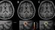
Methods: We analysed T1 and T2 relaxometry maps from a longitudinal cohort of MS patients. Lesions were classified as enlarging, shrinking, new or stable based on their longitudinal volumetric change using a newly developed automated technique. Voxelwise deviations were computed as z-scores by comparing individual patient data to T1, T2 and T2/T1 normative values from healthy subjects. We studied the distribution of microstructural properties inside lesions and within perilesional tissue.
Results and conclusions: Stable lesions exhibited the highest T1 and T2 z-scores in lesion tissue, while the lowest values were observed for new lesions. Shrinking lesions presented the highest T1 z-scores in the first perilesional ring while enlarging lesions showed the highest T2 z-scores in the same region. Finally, a classification model was trained to predict the longitudinal lesion type based on microstructural metrics and feature importance was assessed. Z-scores estimated in lesion and perilesional tissue from T1, T2 and T2/T1 quantitative maps carry discriminative and complementary information to classify longitudinal lesion phenotypes, hence suggesting that multiparametric MRI approaches are essential for a better understanding of the pathophysiological mechanisms underlying disease activity in MS lesions.
期刊介绍:
The Journal of Neurology is an international peer-reviewed journal which provides a source for publishing original communications and reviews on clinical neurology covering the whole field.
In addition, Letters to the Editors serve as a forum for clinical cases and the exchange of ideas which highlight important new findings. A section on Neurological progress serves to summarise the major findings in certain fields of neurology. Commentaries on new developments in clinical neuroscience, which may be commissioned or submitted, are published as editorials.
Every neurologist interested in the current diagnosis and treatment of neurological disorders needs access to the information contained in this valuable journal.

 求助内容:
求助内容: 应助结果提醒方式:


