{"title":"右上肢软组织血管瘤,伴有骨内扩展和继发性血管内乳头状内皮增生。","authors":"Rachel Bass, Gene Siegal, Apoorva Kotha, Yulia Melenevksy","doi":"10.1007/s00256-024-04727-y","DOIUrl":null,"url":null,"abstract":"<p><p>Intravascular papillary endothelial hyperplasia (IPEH), also known as Masson's tumor, is an uncommon exuberant form of organizing thrombus that may occur within a vessel, vascular tumor, or hematoma and may change the imaging appearance to mimic an aggressive process. It must be distinguished pathologically from angiosarcoma. They have been most commonly reported within superficial soft tissue tumors, and rapid growth and effect on bone are rarely described. We present a case of a patient with a soft tissue hemangioma with IPEH with intraosseous extension that presented with a pathologic fracture of her right humerus with an aggressive appearing osseous lesion. CT and MRI demonstrated a multifocal ill-defined soft tissue mass throughout the right upper extremity with underlying cortical tunneling and scalloping of the proximal humerus. Similar imaging findings were also present in the distal humerus and ipsilateral scapula and evolved during her hospitalization. Following percutaneous biopsy revealing hemangioma with features of papillary endothelial hyperplasia with intraosseous extension, the patient died in the ICU secondary to unrelated septic shock. Diagnosis was confirmed at autopsy. Primary and secondary IPEH have been generally characterized as well-defined solitary masses, most often in the superficial soft tissues. This case of a deep soft tissue hemangioma with type II IPEH, intraosseous extension, and imaging findings of regional multicompartmental involvement is very unusual. Reporting of this case in the literature should be beneficial for pathologic correlation with similar confounding masses as well as propose a possible mechanism for intraosseous extension of soft tissue hemangiomas.</p>","PeriodicalId":21783,"journal":{"name":"Skeletal Radiology","volume":" ","pages":"619-625"},"PeriodicalIF":1.9000,"publicationDate":"2025-03-01","publicationTypes":"Journal Article","fieldsOfStudy":null,"isOpenAccess":false,"openAccessPdf":"https://www.ncbi.nlm.nih.gov/pmc/articles/PMC11769867/pdf/","citationCount":"0","resultStr":"{\"title\":\"Soft tissue hemangioma of the right upper extremity with intraosseous extension and secondary intravascular papillary endothelial hyperplasia.\",\"authors\":\"Rachel Bass, Gene Siegal, Apoorva Kotha, Yulia Melenevksy\",\"doi\":\"10.1007/s00256-024-04727-y\",\"DOIUrl\":null,\"url\":null,\"abstract\":\"<p><p>Intravascular papillary endothelial hyperplasia (IPEH), also known as Masson's tumor, is an uncommon exuberant form of organizing thrombus that may occur within a vessel, vascular tumor, or hematoma and may change the imaging appearance to mimic an aggressive process. It must be distinguished pathologically from angiosarcoma. They have been most commonly reported within superficial soft tissue tumors, and rapid growth and effect on bone are rarely described. We present a case of a patient with a soft tissue hemangioma with IPEH with intraosseous extension that presented with a pathologic fracture of her right humerus with an aggressive appearing osseous lesion. CT and MRI demonstrated a multifocal ill-defined soft tissue mass throughout the right upper extremity with underlying cortical tunneling and scalloping of the proximal humerus. Similar imaging findings were also present in the distal humerus and ipsilateral scapula and evolved during her hospitalization. Following percutaneous biopsy revealing hemangioma with features of papillary endothelial hyperplasia with intraosseous extension, the patient died in the ICU secondary to unrelated septic shock. Diagnosis was confirmed at autopsy. Primary and secondary IPEH have been generally characterized as well-defined solitary masses, most often in the superficial soft tissues. This case of a deep soft tissue hemangioma with type II IPEH, intraosseous extension, and imaging findings of regional multicompartmental involvement is very unusual. Reporting of this case in the literature should be beneficial for pathologic correlation with similar confounding masses as well as propose a possible mechanism for intraosseous extension of soft tissue hemangiomas.</p>\",\"PeriodicalId\":21783,\"journal\":{\"name\":\"Skeletal Radiology\",\"volume\":\" \",\"pages\":\"619-625\"},\"PeriodicalIF\":1.9000,\"publicationDate\":\"2025-03-01\",\"publicationTypes\":\"Journal Article\",\"fieldsOfStudy\":null,\"isOpenAccess\":false,\"openAccessPdf\":\"https://www.ncbi.nlm.nih.gov/pmc/articles/PMC11769867/pdf/\",\"citationCount\":\"0\",\"resultStr\":null,\"platform\":\"Semanticscholar\",\"paperid\":null,\"PeriodicalName\":\"Skeletal Radiology\",\"FirstCategoryId\":\"3\",\"ListUrlMain\":\"https://doi.org/10.1007/s00256-024-04727-y\",\"RegionNum\":3,\"RegionCategory\":\"医学\",\"ArticlePicture\":[],\"TitleCN\":null,\"AbstractTextCN\":null,\"PMCID\":null,\"EPubDate\":\"2024/7/12 0:00:00\",\"PubModel\":\"Epub\",\"JCR\":\"Q2\",\"JCRName\":\"ORTHOPEDICS\",\"Score\":null,\"Total\":0}","platform":"Semanticscholar","paperid":null,"PeriodicalName":"Skeletal Radiology","FirstCategoryId":"3","ListUrlMain":"https://doi.org/10.1007/s00256-024-04727-y","RegionNum":3,"RegionCategory":"医学","ArticlePicture":[],"TitleCN":null,"AbstractTextCN":null,"PMCID":null,"EPubDate":"2024/7/12 0:00:00","PubModel":"Epub","JCR":"Q2","JCRName":"ORTHOPEDICS","Score":null,"Total":0}
引用次数: 0
摘要
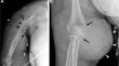
血管内乳头状内皮增生症(IPEH)又称马森氏瘤,是一种不常见的增生性组织血栓,可能发生在血管、血管瘤或血肿内,并可能改变成像外观,模仿侵袭性过程。病理上必须将其与血管肉瘤区分开来。最常见的报道是在浅表软组织肿瘤内发生,很少有快速生长并影响骨骼的描述。我们介绍了一例患有软组织血管瘤并伴有骨内扩展的 IPEH 的患者,她的右肱骨出现病理性骨折,并伴有侵袭性骨病变。CT和磁共振成像显示,患者右上肢有多灶性界限不清的软组织肿块,肱骨近端皮质下有隧道和扇形结构。肱骨远端和同侧肩胛骨也有类似的成像结果,并在住院期间不断发展。经皮穿刺活检显示患者患有血管瘤,并伴有乳头状内皮增生和骨内扩展,随后患者在重症监护室死于与此无关的脓毒性休克。尸检证实了诊断结果。原发性和继发性 IPEH 通常表现为界限清楚的单发肿块,多见于表层软组织。这例深部软组织血管瘤伴有 II 型 IPEH、骨内扩展和区域性多室受累的影像学发现非常罕见。该病例的文献报道将有助于与类似的混杂肿块进行病理关联,并提出软组织血管瘤骨内扩展的可能机制。
Soft tissue hemangioma of the right upper extremity with intraosseous extension and secondary intravascular papillary endothelial hyperplasia.
Intravascular papillary endothelial hyperplasia (IPEH), also known as Masson's tumor, is an uncommon exuberant form of organizing thrombus that may occur within a vessel, vascular tumor, or hematoma and may change the imaging appearance to mimic an aggressive process. It must be distinguished pathologically from angiosarcoma. They have been most commonly reported within superficial soft tissue tumors, and rapid growth and effect on bone are rarely described. We present a case of a patient with a soft tissue hemangioma with IPEH with intraosseous extension that presented with a pathologic fracture of her right humerus with an aggressive appearing osseous lesion. CT and MRI demonstrated a multifocal ill-defined soft tissue mass throughout the right upper extremity with underlying cortical tunneling and scalloping of the proximal humerus. Similar imaging findings were also present in the distal humerus and ipsilateral scapula and evolved during her hospitalization. Following percutaneous biopsy revealing hemangioma with features of papillary endothelial hyperplasia with intraosseous extension, the patient died in the ICU secondary to unrelated septic shock. Diagnosis was confirmed at autopsy. Primary and secondary IPEH have been generally characterized as well-defined solitary masses, most often in the superficial soft tissues. This case of a deep soft tissue hemangioma with type II IPEH, intraosseous extension, and imaging findings of regional multicompartmental involvement is very unusual. Reporting of this case in the literature should be beneficial for pathologic correlation with similar confounding masses as well as propose a possible mechanism for intraosseous extension of soft tissue hemangiomas.
期刊介绍:
Skeletal Radiology provides a forum for the dissemination of current knowledge and information dealing with disorders of the musculoskeletal system including the spine. While emphasizing the radiological aspects of the many varied skeletal abnormalities, the journal also adopts an interdisciplinary approach, reflecting the membership of the International Skeletal Society. Thus, the anatomical, pathological, physiological, clinical, metabolic and epidemiological aspects of the many entities affecting the skeleton receive appropriate consideration.
This is the Journal of the International Skeletal Society and the Official Journal of the Society of Skeletal Radiology and the Australasian Musculoskelelal Imaging Group.

 求助内容:
求助内容: 应助结果提醒方式:


