{"title":"无创肝脏脂肪定量:多回波 Dixon 能帮上忙吗?","authors":"Akarshi Gupta, Rashmi Dixit, Anjali Prakash","doi":"10.1590/0100-3984.2023.0125","DOIUrl":null,"url":null,"abstract":"<p><strong>Objective: </strong>To evaluate the diagnostic accuracy of multi-echo Dixon magnetic resonance imaging (MRI) in hepatic fat quantification, in comparison with that of magnetic resonance spectroscopy (MRS), on 3.0-T MRI.</p><p><strong>Materials and methods: </strong>Fifty-five adults with no known liver disease underwent MRI in a 3.0-T scanner for determination of the hepatic fat fraction, with two techniques: multi-echo Dixon, in a manually drawn region of interest (ROI) and in the entire liver parenchyma (automated segmentation); and MRS. The diagnostic accuracy and cutoff value for multi-echo Dixon were determined, with MRS being used as the reference standard.</p><p><strong>Results: </strong>The mean fat fraction obtained by multi-echo Dixon in the manually drawn ROI and in the entire liver was 5.2 ± 5.8% and 6.6 ± 5.2%, respectively, whereas the mean hepatic fat fraction obtained by MRS was 5.7 ± 6.4%. A very strong positive correlation and good agreement were observed between MRS and multi-echo Dixon, for the ROI (r = 0.988, r<sup>2</sup> = 0.978, <i>p</i> < 0.001) and for the entire liver parenchyma (r = 0.960, r<sup>2</sup> = 0.922, <i>p</i> < 0.001). A moderate positive correlation was observed between the hepatic fat fraction and body mass index of the participants, regardless of the fat estimation technique employed.</p><p><strong>Conclusion: </strong>For hepatic fat quantification, multi-echo Dixon MRI demonstrated a very strong positive correlation and good agreement with MRS (often considered the gold-standard noninvasive technique). Because multi-echo Dixon MRI is more readily available than is MRS, it can be used as a rapid tool for hepatic fat quantification, especially when the hepatic fat distribution is not homogeneous.</p>","PeriodicalId":20842,"journal":{"name":"Radiologia Brasileira","volume":"57 ","pages":"e20230125"},"PeriodicalIF":0.0000,"publicationDate":"2024-05-07","publicationTypes":"Journal Article","fieldsOfStudy":null,"isOpenAccess":false,"openAccessPdf":"https://www.ncbi.nlm.nih.gov/pmc/articles/PMC11235074/pdf/","citationCount":"0","resultStr":"{\"title\":\"Non-invasive hepatic fat quantification: Can multi-echo Dixon help?\",\"authors\":\"Akarshi Gupta, Rashmi Dixit, Anjali Prakash\",\"doi\":\"10.1590/0100-3984.2023.0125\",\"DOIUrl\":null,\"url\":null,\"abstract\":\"<p><strong>Objective: </strong>To evaluate the diagnostic accuracy of multi-echo Dixon magnetic resonance imaging (MRI) in hepatic fat quantification, in comparison with that of magnetic resonance spectroscopy (MRS), on 3.0-T MRI.</p><p><strong>Materials and methods: </strong>Fifty-five adults with no known liver disease underwent MRI in a 3.0-T scanner for determination of the hepatic fat fraction, with two techniques: multi-echo Dixon, in a manually drawn region of interest (ROI) and in the entire liver parenchyma (automated segmentation); and MRS. The diagnostic accuracy and cutoff value for multi-echo Dixon were determined, with MRS being used as the reference standard.</p><p><strong>Results: </strong>The mean fat fraction obtained by multi-echo Dixon in the manually drawn ROI and in the entire liver was 5.2 ± 5.8% and 6.6 ± 5.2%, respectively, whereas the mean hepatic fat fraction obtained by MRS was 5.7 ± 6.4%. A very strong positive correlation and good agreement were observed between MRS and multi-echo Dixon, for the ROI (r = 0.988, r<sup>2</sup> = 0.978, <i>p</i> < 0.001) and for the entire liver parenchyma (r = 0.960, r<sup>2</sup> = 0.922, <i>p</i> < 0.001). A moderate positive correlation was observed between the hepatic fat fraction and body mass index of the participants, regardless of the fat estimation technique employed.</p><p><strong>Conclusion: </strong>For hepatic fat quantification, multi-echo Dixon MRI demonstrated a very strong positive correlation and good agreement with MRS (often considered the gold-standard noninvasive technique). Because multi-echo Dixon MRI is more readily available than is MRS, it can be used as a rapid tool for hepatic fat quantification, especially when the hepatic fat distribution is not homogeneous.</p>\",\"PeriodicalId\":20842,\"journal\":{\"name\":\"Radiologia Brasileira\",\"volume\":\"57 \",\"pages\":\"e20230125\"},\"PeriodicalIF\":0.0000,\"publicationDate\":\"2024-05-07\",\"publicationTypes\":\"Journal Article\",\"fieldsOfStudy\":null,\"isOpenAccess\":false,\"openAccessPdf\":\"https://www.ncbi.nlm.nih.gov/pmc/articles/PMC11235074/pdf/\",\"citationCount\":\"0\",\"resultStr\":null,\"platform\":\"Semanticscholar\",\"paperid\":null,\"PeriodicalName\":\"Radiologia Brasileira\",\"FirstCategoryId\":\"1085\",\"ListUrlMain\":\"https://doi.org/10.1590/0100-3984.2023.0125\",\"RegionNum\":0,\"RegionCategory\":null,\"ArticlePicture\":[],\"TitleCN\":null,\"AbstractTextCN\":null,\"PMCID\":null,\"EPubDate\":\"2024/1/1 0:00:00\",\"PubModel\":\"eCollection\",\"JCR\":\"Q3\",\"JCRName\":\"Medicine\",\"Score\":null,\"Total\":0}","platform":"Semanticscholar","paperid":null,"PeriodicalName":"Radiologia Brasileira","FirstCategoryId":"1085","ListUrlMain":"https://doi.org/10.1590/0100-3984.2023.0125","RegionNum":0,"RegionCategory":null,"ArticlePicture":[],"TitleCN":null,"AbstractTextCN":null,"PMCID":null,"EPubDate":"2024/1/1 0:00:00","PubModel":"eCollection","JCR":"Q3","JCRName":"Medicine","Score":null,"Total":0}
Non-invasive hepatic fat quantification: Can multi-echo Dixon help?
Objective: To evaluate the diagnostic accuracy of multi-echo Dixon magnetic resonance imaging (MRI) in hepatic fat quantification, in comparison with that of magnetic resonance spectroscopy (MRS), on 3.0-T MRI.
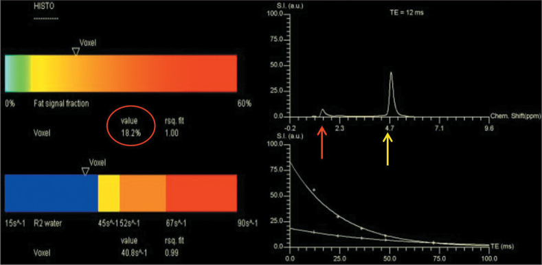
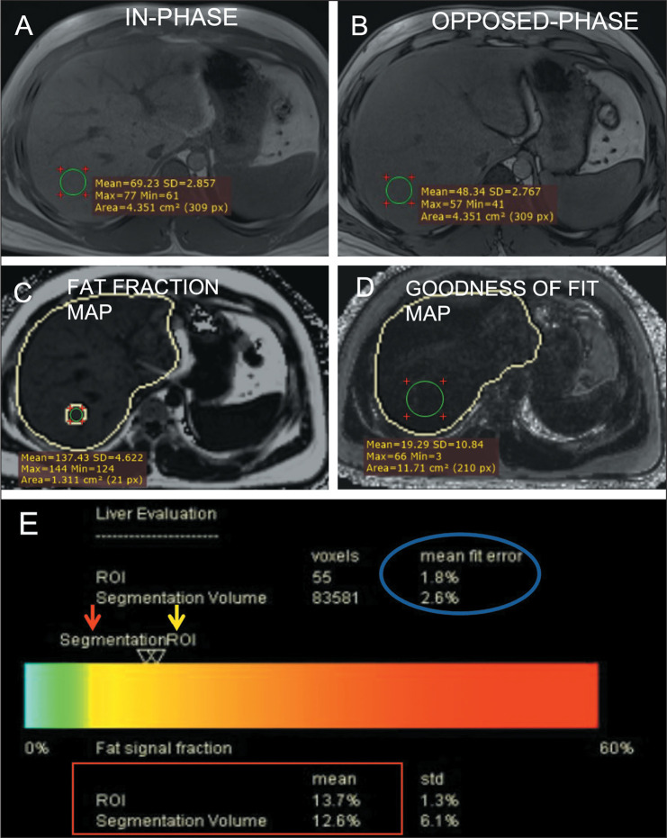
Materials and methods: Fifty-five adults with no known liver disease underwent MRI in a 3.0-T scanner for determination of the hepatic fat fraction, with two techniques: multi-echo Dixon, in a manually drawn region of interest (ROI) and in the entire liver parenchyma (automated segmentation); and MRS. The diagnostic accuracy and cutoff value for multi-echo Dixon were determined, with MRS being used as the reference standard.
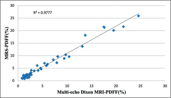
Results: The mean fat fraction obtained by multi-echo Dixon in the manually drawn ROI and in the entire liver was 5.2 ± 5.8% and 6.6 ± 5.2%, respectively, whereas the mean hepatic fat fraction obtained by MRS was 5.7 ± 6.4%. A very strong positive correlation and good agreement were observed between MRS and multi-echo Dixon, for the ROI (r = 0.988, r2 = 0.978, p < 0.001) and for the entire liver parenchyma (r = 0.960, r2 = 0.922, p < 0.001). A moderate positive correlation was observed between the hepatic fat fraction and body mass index of the participants, regardless of the fat estimation technique employed.
Conclusion: For hepatic fat quantification, multi-echo Dixon MRI demonstrated a very strong positive correlation and good agreement with MRS (often considered the gold-standard noninvasive technique). Because multi-echo Dixon MRI is more readily available than is MRS, it can be used as a rapid tool for hepatic fat quantification, especially when the hepatic fat distribution is not homogeneous.

 求助内容:
求助内容: 应助结果提醒方式:


