{"title":"利用微图案图像进行深度学习,量化早期分化的人类诱导多能干细胞的空间图案和形成过程。","authors":"Slo-Li Chu, Kuniya Abe, Hideo Yokota, Dooseon Cho, Yohei Hayashi, Ming-Dar Tsai","doi":"10.1111/jmi.13346","DOIUrl":null,"url":null,"abstract":"<p>Micropatterning is reliable method for quantifying pluripotency of human-induced pluripotent stem cells (hiPSCs) that differentiate to form a spatial pattern of sorted, ordered and nonoverlapped three germ layers on the micropattern. In this study, we propose a deep learning method to quantify spatial patterning of the germ layers in the early differentiation stage of hiPSCs using micropattern images. We propose decoding and encoding U-net structures learning labelled Hoechst (DNA-stained) hiPSC regions with corresponding Hoechst and bright-field micropattern images to segment hiPSCs on Hoechst or bright-field images. We also propose a U-net structure to extract extraembryonic regions on a micropattern, and an algorithm to compares intensities of the fluorescence images staining respective germ-layer cells and extract their regions. The proposed method thus can quantify the pluripotency of a hiPSC line with spatial patterning including cell numbers, areas and distributions of germ-layer and extraembryonic cells on a micropattern, and reveal the formation process of hiPSCs and germ layers in the early differentiation stage by segmenting live-cell bright-field images. In our assay, the cell-number accuracy achieved 86% and 85%, and the cell region accuracy 89% and 81% for segmenting Hoechst and bright-field micropattern images, respectively. Applications to micropattern images of multiple hiPSC lines, micropattern sizes, groups of markers, living and fixed cells show the proposed method can be expected to be a useful protocol and tool to quantify pluripotency of a new hiPSC line before providing it to the scientific community.</p>","PeriodicalId":16484,"journal":{"name":"Journal of microscopy","volume":"296 1","pages":"79-93"},"PeriodicalIF":1.9000,"publicationDate":"2024-07-12","publicationTypes":"Journal Article","fieldsOfStudy":null,"isOpenAccess":false,"openAccessPdf":"https://onlinelibrary.wiley.com/doi/epdf/10.1111/jmi.13346","citationCount":"0","resultStr":"{\"title\":\"Deep learning for quantifying spatial patterning and formation process of early differentiated human-induced pluripotent stem cells with micropattern images\",\"authors\":\"Slo-Li Chu, Kuniya Abe, Hideo Yokota, Dooseon Cho, Yohei Hayashi, Ming-Dar Tsai\",\"doi\":\"10.1111/jmi.13346\",\"DOIUrl\":null,\"url\":null,\"abstract\":\"<p>Micropatterning is reliable method for quantifying pluripotency of human-induced pluripotent stem cells (hiPSCs) that differentiate to form a spatial pattern of sorted, ordered and nonoverlapped three germ layers on the micropattern. In this study, we propose a deep learning method to quantify spatial patterning of the germ layers in the early differentiation stage of hiPSCs using micropattern images. We propose decoding and encoding U-net structures learning labelled Hoechst (DNA-stained) hiPSC regions with corresponding Hoechst and bright-field micropattern images to segment hiPSCs on Hoechst or bright-field images. We also propose a U-net structure to extract extraembryonic regions on a micropattern, and an algorithm to compares intensities of the fluorescence images staining respective germ-layer cells and extract their regions. The proposed method thus can quantify the pluripotency of a hiPSC line with spatial patterning including cell numbers, areas and distributions of germ-layer and extraembryonic cells on a micropattern, and reveal the formation process of hiPSCs and germ layers in the early differentiation stage by segmenting live-cell bright-field images. In our assay, the cell-number accuracy achieved 86% and 85%, and the cell region accuracy 89% and 81% for segmenting Hoechst and bright-field micropattern images, respectively. Applications to micropattern images of multiple hiPSC lines, micropattern sizes, groups of markers, living and fixed cells show the proposed method can be expected to be a useful protocol and tool to quantify pluripotency of a new hiPSC line before providing it to the scientific community.</p>\",\"PeriodicalId\":16484,\"journal\":{\"name\":\"Journal of microscopy\",\"volume\":\"296 1\",\"pages\":\"79-93\"},\"PeriodicalIF\":1.9000,\"publicationDate\":\"2024-07-12\",\"publicationTypes\":\"Journal Article\",\"fieldsOfStudy\":null,\"isOpenAccess\":false,\"openAccessPdf\":\"https://onlinelibrary.wiley.com/doi/epdf/10.1111/jmi.13346\",\"citationCount\":\"0\",\"resultStr\":null,\"platform\":\"Semanticscholar\",\"paperid\":null,\"PeriodicalName\":\"Journal of microscopy\",\"FirstCategoryId\":\"5\",\"ListUrlMain\":\"https://onlinelibrary.wiley.com/doi/10.1111/jmi.13346\",\"RegionNum\":4,\"RegionCategory\":\"工程技术\",\"ArticlePicture\":[],\"TitleCN\":null,\"AbstractTextCN\":null,\"PMCID\":null,\"EPubDate\":\"\",\"PubModel\":\"\",\"JCR\":\"Q3\",\"JCRName\":\"MICROSCOPY\",\"Score\":null,\"Total\":0}","platform":"Semanticscholar","paperid":null,"PeriodicalName":"Journal of microscopy","FirstCategoryId":"5","ListUrlMain":"https://onlinelibrary.wiley.com/doi/10.1111/jmi.13346","RegionNum":4,"RegionCategory":"工程技术","ArticlePicture":[],"TitleCN":null,"AbstractTextCN":null,"PMCID":null,"EPubDate":"","PubModel":"","JCR":"Q3","JCRName":"MICROSCOPY","Score":null,"Total":0}
Deep learning for quantifying spatial patterning and formation process of early differentiated human-induced pluripotent stem cells with micropattern images
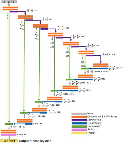
Micropatterning is reliable method for quantifying pluripotency of human-induced pluripotent stem cells (hiPSCs) that differentiate to form a spatial pattern of sorted, ordered and nonoverlapped three germ layers on the micropattern. In this study, we propose a deep learning method to quantify spatial patterning of the germ layers in the early differentiation stage of hiPSCs using micropattern images. We propose decoding and encoding U-net structures learning labelled Hoechst (DNA-stained) hiPSC regions with corresponding Hoechst and bright-field micropattern images to segment hiPSCs on Hoechst or bright-field images. We also propose a U-net structure to extract extraembryonic regions on a micropattern, and an algorithm to compares intensities of the fluorescence images staining respective germ-layer cells and extract their regions. The proposed method thus can quantify the pluripotency of a hiPSC line with spatial patterning including cell numbers, areas and distributions of germ-layer and extraembryonic cells on a micropattern, and reveal the formation process of hiPSCs and germ layers in the early differentiation stage by segmenting live-cell bright-field images. In our assay, the cell-number accuracy achieved 86% and 85%, and the cell region accuracy 89% and 81% for segmenting Hoechst and bright-field micropattern images, respectively. Applications to micropattern images of multiple hiPSC lines, micropattern sizes, groups of markers, living and fixed cells show the proposed method can be expected to be a useful protocol and tool to quantify pluripotency of a new hiPSC line before providing it to the scientific community.
期刊介绍:
The Journal of Microscopy is the oldest journal dedicated to the science of microscopy and the only peer-reviewed publication of the Royal Microscopical Society. It publishes papers that report on the very latest developments in microscopy such as advances in microscopy techniques or novel areas of application. The Journal does not seek to publish routine applications of microscopy or specimen preparation even though the submission may otherwise have a high scientific merit.
The scope covers research in the physical and biological sciences and covers imaging methods using light, electrons, X-rays and other radiations as well as atomic force and near field techniques. Interdisciplinary research is welcome. Papers pertaining to microscopy are also welcomed on optical theory, spectroscopy, novel specimen preparation and manipulation methods and image recording, processing and analysis including dynamic analysis of living specimens.
Publication types include full papers, hot topic fast tracked communications and review articles. Authors considering submitting a review article should contact the editorial office first.

 求助内容:
求助内容: 应助结果提醒方式:


