Nicole Tonin Iplinsky, Luiz Gonzaga Gandini Junior, Alyssa Schiavon Gandini, Ana Thais Bagatini, Pedro Henrique José de Oliveira, Paula Cristina Henriques da Silva, Ary Santos-Pinto
{"title":"恒牙珐琅质厚度的 X 射线评估:相关性和适用性。","authors":"Nicole Tonin Iplinsky, Luiz Gonzaga Gandini Junior, Alyssa Schiavon Gandini, Ana Thais Bagatini, Pedro Henrique José de Oliveira, Paula Cristina Henriques da Silva, Ary Santos-Pinto","doi":"10.1590/2177-6709.29.3.e242422.oar","DOIUrl":null,"url":null,"abstract":"<p><strong>Objective: </strong>This descriptive observational study aimed to determine clinically relevant and applicable data of enamel thickness (ETH), considering the mesio-distal differences of anterior and posterior permanent teeth and their relationships.</p><p><strong>Material and methods: </strong>The sample consisted of right-sided standardized radiographs of 34 individuals (21 females and 13 males), aged between 13 and 24 (average 16) years, with all permanent teeth intact and without crowding. Four periapical and four interproximal radiographs were obtained and digitized. ETH measurements (mesial to distal contact points at the dentin-enamel junction) were performed after correction for radiographic image magnification. The Students' t-test was applied to the differences between paired means, with the Pearson correlation to evaluate the correlation between them.</p><p><strong>Results: </strong>The mesial and distal ETH increased from the anterior to the posterior teeth. Incisor ETH ranged between 0.60 and 0.84 mm. Canines, premolars, and molars were more than 1.0 mm thick, and molar enamel reached values between 1.26 and 1.44 mm.</p><p><strong>Conclusion: </strong>Distal ETH was significantly greater than the mesial ETH, and progressively thicker from the anterior to posterior teeth. Interproximal reduction (IPR) of the lower central and upper lateral incisors should be avoided, reduced, or performed on their distal surfaces. There is a positive and significant correlation between ETH and the mesial and distal surfaces of the teeth. Periapical radiographs and evaluation of the remaining ETH are necessary in cases of retreatment. The location and number of tooth size discrepancies should be considered in treatment planning and appropriately compensated with IPR.</p>","PeriodicalId":38720,"journal":{"name":"Dental Press Journal of Orthodontics","volume":"29 3","pages":"e242422"},"PeriodicalIF":0.0000,"publicationDate":"2024-07-08","publicationTypes":"Journal Article","fieldsOfStudy":null,"isOpenAccess":false,"openAccessPdf":"https://www.ncbi.nlm.nih.gov/pmc/articles/PMC11235574/pdf/","citationCount":"0","resultStr":"{\"title\":\"Radiographic evaluation of enamel thickness of permanent teeth: relevance and applicability.\",\"authors\":\"Nicole Tonin Iplinsky, Luiz Gonzaga Gandini Junior, Alyssa Schiavon Gandini, Ana Thais Bagatini, Pedro Henrique José de Oliveira, Paula Cristina Henriques da Silva, Ary Santos-Pinto\",\"doi\":\"10.1590/2177-6709.29.3.e242422.oar\",\"DOIUrl\":null,\"url\":null,\"abstract\":\"<p><strong>Objective: </strong>This descriptive observational study aimed to determine clinically relevant and applicable data of enamel thickness (ETH), considering the mesio-distal differences of anterior and posterior permanent teeth and their relationships.</p><p><strong>Material and methods: </strong>The sample consisted of right-sided standardized radiographs of 34 individuals (21 females and 13 males), aged between 13 and 24 (average 16) years, with all permanent teeth intact and without crowding. Four periapical and four interproximal radiographs were obtained and digitized. ETH measurements (mesial to distal contact points at the dentin-enamel junction) were performed after correction for radiographic image magnification. The Students' t-test was applied to the differences between paired means, with the Pearson correlation to evaluate the correlation between them.</p><p><strong>Results: </strong>The mesial and distal ETH increased from the anterior to the posterior teeth. Incisor ETH ranged between 0.60 and 0.84 mm. Canines, premolars, and molars were more than 1.0 mm thick, and molar enamel reached values between 1.26 and 1.44 mm.</p><p><strong>Conclusion: </strong>Distal ETH was significantly greater than the mesial ETH, and progressively thicker from the anterior to posterior teeth. Interproximal reduction (IPR) of the lower central and upper lateral incisors should be avoided, reduced, or performed on their distal surfaces. There is a positive and significant correlation between ETH and the mesial and distal surfaces of the teeth. Periapical radiographs and evaluation of the remaining ETH are necessary in cases of retreatment. The location and number of tooth size discrepancies should be considered in treatment planning and appropriately compensated with IPR.</p>\",\"PeriodicalId\":38720,\"journal\":{\"name\":\"Dental Press Journal of Orthodontics\",\"volume\":\"29 3\",\"pages\":\"e242422\"},\"PeriodicalIF\":0.0000,\"publicationDate\":\"2024-07-08\",\"publicationTypes\":\"Journal Article\",\"fieldsOfStudy\":null,\"isOpenAccess\":false,\"openAccessPdf\":\"https://www.ncbi.nlm.nih.gov/pmc/articles/PMC11235574/pdf/\",\"citationCount\":\"0\",\"resultStr\":null,\"platform\":\"Semanticscholar\",\"paperid\":null,\"PeriodicalName\":\"Dental Press Journal of Orthodontics\",\"FirstCategoryId\":\"1085\",\"ListUrlMain\":\"https://doi.org/10.1590/2177-6709.29.3.e242422.oar\",\"RegionNum\":0,\"RegionCategory\":null,\"ArticlePicture\":[],\"TitleCN\":null,\"AbstractTextCN\":null,\"PMCID\":null,\"EPubDate\":\"2024/1/1 0:00:00\",\"PubModel\":\"eCollection\",\"JCR\":\"Q2\",\"JCRName\":\"Medicine\",\"Score\":null,\"Total\":0}","platform":"Semanticscholar","paperid":null,"PeriodicalName":"Dental Press Journal of Orthodontics","FirstCategoryId":"1085","ListUrlMain":"https://doi.org/10.1590/2177-6709.29.3.e242422.oar","RegionNum":0,"RegionCategory":null,"ArticlePicture":[],"TitleCN":null,"AbstractTextCN":null,"PMCID":null,"EPubDate":"2024/1/1 0:00:00","PubModel":"eCollection","JCR":"Q2","JCRName":"Medicine","Score":null,"Total":0}
Radiographic evaluation of enamel thickness of permanent teeth: relevance and applicability.
Objective: This descriptive observational study aimed to determine clinically relevant and applicable data of enamel thickness (ETH), considering the mesio-distal differences of anterior and posterior permanent teeth and their relationships.
Material and methods: The sample consisted of right-sided standardized radiographs of 34 individuals (21 females and 13 males), aged between 13 and 24 (average 16) years, with all permanent teeth intact and without crowding. Four periapical and four interproximal radiographs were obtained and digitized. ETH measurements (mesial to distal contact points at the dentin-enamel junction) were performed after correction for radiographic image magnification. The Students' t-test was applied to the differences between paired means, with the Pearson correlation to evaluate the correlation between them.
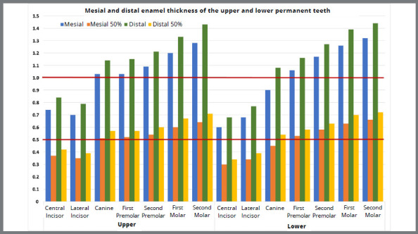
Results: The mesial and distal ETH increased from the anterior to the posterior teeth. Incisor ETH ranged between 0.60 and 0.84 mm. Canines, premolars, and molars were more than 1.0 mm thick, and molar enamel reached values between 1.26 and 1.44 mm.
Conclusion: Distal ETH was significantly greater than the mesial ETH, and progressively thicker from the anterior to posterior teeth. Interproximal reduction (IPR) of the lower central and upper lateral incisors should be avoided, reduced, or performed on their distal surfaces. There is a positive and significant correlation between ETH and the mesial and distal surfaces of the teeth. Periapical radiographs and evaluation of the remaining ETH are necessary in cases of retreatment. The location and number of tooth size discrepancies should be considered in treatment planning and appropriately compensated with IPR.
期刊介绍:
The Dental Press Journal of Orthodontics publishes scientific research articles, significant reviews, clinical and technical case reports, brief communications, and other materials related to Orthodontics and Facial Orthopedics.

 求助内容:
求助内容: 应助结果提醒方式:


