Alline Birra Nolasco Fernandes, Luíza Trindade Vilela, Taísa Figueiredo Chagas, Antônio Carlos de Oliveira Ruellas, Cláudia Trindade Mattos, Margareth Maria Gomes de Souza
{"title":"下颌前突矫形手术会导致髁突改变吗?病例系列分析。","authors":"Alline Birra Nolasco Fernandes, Luíza Trindade Vilela, Taísa Figueiredo Chagas, Antônio Carlos de Oliveira Ruellas, Cláudia Trindade Mattos, Margareth Maria Gomes de Souza","doi":"10.1590/2177-6709.29.3.e2423261.oar","DOIUrl":null,"url":null,"abstract":"<p><strong>Introduction: </strong>Mandibular advancement surgery corrects bone bases while establishing patients' functional and aesthetic rehabilitation. However, little is known about the results of this procedure in the structures that make up the stomatognathic system, as the condyles.</p><p><strong>Objective: </strong>This study aimed to evaluate the structural and positional changes of mandibular condyles in ortho-surgical patients who underwent mandibular advancement surgery.</p><p><strong>Material and methods: </strong>A prospective investigation was conducted with cone-beam computed tomography images. Using Dolphin Imaging® software, seven ortho-surgical patients with Angle Class II malocclusion and mandibular deficiency were evaluated. The images assessed were obtained at pre-surgical phase and after, at least, 1 year of the procedure. To study the structural and positional changes of condyles, linear and angular measurements were obtained, and the right and left sides of patients were compared. Descriptive statistical analysis was performed and, in order to verify possible significant differences, normality tests (Kolmogorov-Smirnov) were applied, followed by a paired t-test to define significance.</p><p><strong>Results: </strong>For all measures evaluated in this study, no statistically significant differences were found.</p><p><strong>Conclusion: </strong>The ortho-surgical procedure performed did not change the structure and position of the condyles of patients who underwent surgical mandibular advancement. Right and left mandibular condyles behaved similarly, suggesting stability and condylar adaptation after surgery.</p>","PeriodicalId":38720,"journal":{"name":"Dental Press Journal of Orthodontics","volume":"29 3","pages":"e2423261"},"PeriodicalIF":0.0000,"publicationDate":"2024-07-08","publicationTypes":"Journal Article","fieldsOfStudy":null,"isOpenAccess":false,"openAccessPdf":"https://www.ncbi.nlm.nih.gov/pmc/articles/PMC11235570/pdf/","citationCount":"0","resultStr":"{\"title\":\"Does mandibular advancement ortho-surgical procedure cause condyle changes? A case-series analysis.\",\"authors\":\"Alline Birra Nolasco Fernandes, Luíza Trindade Vilela, Taísa Figueiredo Chagas, Antônio Carlos de Oliveira Ruellas, Cláudia Trindade Mattos, Margareth Maria Gomes de Souza\",\"doi\":\"10.1590/2177-6709.29.3.e2423261.oar\",\"DOIUrl\":null,\"url\":null,\"abstract\":\"<p><strong>Introduction: </strong>Mandibular advancement surgery corrects bone bases while establishing patients' functional and aesthetic rehabilitation. However, little is known about the results of this procedure in the structures that make up the stomatognathic system, as the condyles.</p><p><strong>Objective: </strong>This study aimed to evaluate the structural and positional changes of mandibular condyles in ortho-surgical patients who underwent mandibular advancement surgery.</p><p><strong>Material and methods: </strong>A prospective investigation was conducted with cone-beam computed tomography images. Using Dolphin Imaging® software, seven ortho-surgical patients with Angle Class II malocclusion and mandibular deficiency were evaluated. The images assessed were obtained at pre-surgical phase and after, at least, 1 year of the procedure. To study the structural and positional changes of condyles, linear and angular measurements were obtained, and the right and left sides of patients were compared. Descriptive statistical analysis was performed and, in order to verify possible significant differences, normality tests (Kolmogorov-Smirnov) were applied, followed by a paired t-test to define significance.</p><p><strong>Results: </strong>For all measures evaluated in this study, no statistically significant differences were found.</p><p><strong>Conclusion: </strong>The ortho-surgical procedure performed did not change the structure and position of the condyles of patients who underwent surgical mandibular advancement. Right and left mandibular condyles behaved similarly, suggesting stability and condylar adaptation after surgery.</p>\",\"PeriodicalId\":38720,\"journal\":{\"name\":\"Dental Press Journal of Orthodontics\",\"volume\":\"29 3\",\"pages\":\"e2423261\"},\"PeriodicalIF\":0.0000,\"publicationDate\":\"2024-07-08\",\"publicationTypes\":\"Journal Article\",\"fieldsOfStudy\":null,\"isOpenAccess\":false,\"openAccessPdf\":\"https://www.ncbi.nlm.nih.gov/pmc/articles/PMC11235570/pdf/\",\"citationCount\":\"0\",\"resultStr\":null,\"platform\":\"Semanticscholar\",\"paperid\":null,\"PeriodicalName\":\"Dental Press Journal of Orthodontics\",\"FirstCategoryId\":\"1085\",\"ListUrlMain\":\"https://doi.org/10.1590/2177-6709.29.3.e2423261.oar\",\"RegionNum\":0,\"RegionCategory\":null,\"ArticlePicture\":[],\"TitleCN\":null,\"AbstractTextCN\":null,\"PMCID\":null,\"EPubDate\":\"2024/1/1 0:00:00\",\"PubModel\":\"eCollection\",\"JCR\":\"Q2\",\"JCRName\":\"Medicine\",\"Score\":null,\"Total\":0}","platform":"Semanticscholar","paperid":null,"PeriodicalName":"Dental Press Journal of Orthodontics","FirstCategoryId":"1085","ListUrlMain":"https://doi.org/10.1590/2177-6709.29.3.e2423261.oar","RegionNum":0,"RegionCategory":null,"ArticlePicture":[],"TitleCN":null,"AbstractTextCN":null,"PMCID":null,"EPubDate":"2024/1/1 0:00:00","PubModel":"eCollection","JCR":"Q2","JCRName":"Medicine","Score":null,"Total":0}
引用次数: 0
摘要
简介下颌骨前移手术在矫正骨基的同时,还能使患者的功能和美观得到康复。然而,人们对构成口颌系统的髁状突结构的手术效果知之甚少:本研究旨在评估接受下颌骨前移手术的矫形外科患者下颌骨髁状突的结构和位置变化:采用锥形束计算机断层扫描图像进行前瞻性研究。使用 Dolphin Imaging® 软件,对七名角Ⅱ类错颌畸形和下颌骨缺损的正畸手术患者进行了评估。所评估的图像是在手术前阶段和手术至少一年后获得的。为了研究髁突的结构和位置变化,对患者的左右两侧进行了线性和角度测量和比较。研究人员进行了描述性统计分析,为了验证可能存在的显著差异,研究人员进行了正态性检验(Kolmogorov-Smirnov),然后进行了配对 t 检验以确定显著性:结果:在本研究评估的所有指标中,均未发现统计学上的显著差异:结论:接受下颌骨前移手术的患者,其髁突的结构和位置并未发生改变。右侧和左侧下颌骨髁突的表现相似,表明手术后髁突的稳定性和适应性。
Does mandibular advancement ortho-surgical procedure cause condyle changes? A case-series analysis.
Introduction: Mandibular advancement surgery corrects bone bases while establishing patients' functional and aesthetic rehabilitation. However, little is known about the results of this procedure in the structures that make up the stomatognathic system, as the condyles.
Objective: This study aimed to evaluate the structural and positional changes of mandibular condyles in ortho-surgical patients who underwent mandibular advancement surgery.
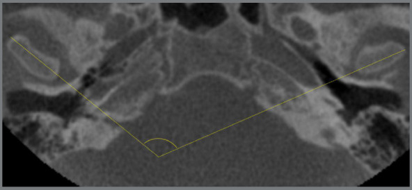
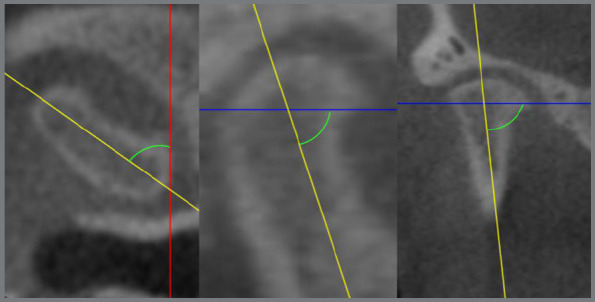
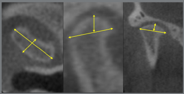
Material and methods: A prospective investigation was conducted with cone-beam computed tomography images. Using Dolphin Imaging® software, seven ortho-surgical patients with Angle Class II malocclusion and mandibular deficiency were evaluated. The images assessed were obtained at pre-surgical phase and after, at least, 1 year of the procedure. To study the structural and positional changes of condyles, linear and angular measurements were obtained, and the right and left sides of patients were compared. Descriptive statistical analysis was performed and, in order to verify possible significant differences, normality tests (Kolmogorov-Smirnov) were applied, followed by a paired t-test to define significance.
Results: For all measures evaluated in this study, no statistically significant differences were found.
Conclusion: The ortho-surgical procedure performed did not change the structure and position of the condyles of patients who underwent surgical mandibular advancement. Right and left mandibular condyles behaved similarly, suggesting stability and condylar adaptation after surgery.
期刊介绍:
The Dental Press Journal of Orthodontics publishes scientific research articles, significant reviews, clinical and technical case reports, brief communications, and other materials related to Orthodontics and Facial Orthopedics.

 求助内容:
求助内容: 应助结果提醒方式:


