Ruoyang Gao, Ling Zhang, Fen Tao, Jun Wang, Guohao Du, Tiqiao Xiao and Biao Deng
{"title":"基于透射 X 射线显微镜的三维 XANES 成像","authors":"Ruoyang Gao, Ling Zhang, Fen Tao, Jun Wang, Guohao Du, Tiqiao Xiao and Biao Deng","doi":"10.1039/D4AN00705K","DOIUrl":null,"url":null,"abstract":"<p >Full-field transmission X-ray microscopy (TXM) in conjunction with X-ray absorption near edge structure (XANES) spectroscopy provides two-dimensional (2D) or three-dimensional (3D) morphological and chemical-specific information within samples at the tens of nanometer scale. This technique has a broad range of applications in materials sciences and battery research. Despite its extensive applicability, 2D XANES imaging is subject to the disadvantage of information overlap when the sample thickness is uneven. 3D XANES imaging combines 3D TXM with XANES to obtain 3D distribution information on chemical states. A 3D XANES imaging method has been established at the Shanghai Synchrotron Radiation Facility (SSRF) and has been used to characterize the structure and chemical state of commercial LiNi<small><sub><em>x</em></sub></small>Co<small><sub><em>y</em></sub></small>Mn<small><sub><em>z</em></sub></small>O<small><sub>2</sub></small> (NCM, <em>x</em> + <em>y</em> + <em>z</em> = 1) battery powder materials. The imaging results provide a visual representation of the 3D chemical state information of the particles with depth resolution, allowing for the direct observation of 3D nickel oxidation. This paper will describe in detail the data acquisition, data processing, quantification and visualization analysis of 3D XANES imaging.</p>","PeriodicalId":63,"journal":{"name":"Analyst","volume":null,"pages":null},"PeriodicalIF":3.6000,"publicationDate":"2024-07-09","publicationTypes":"Journal Article","fieldsOfStudy":null,"isOpenAccess":false,"openAccessPdf":"","citationCount":"0","resultStr":"{\"title\":\"Transmission X-ray microscopy-based three-dimensional XANES imaging\",\"authors\":\"Ruoyang Gao, Ling Zhang, Fen Tao, Jun Wang, Guohao Du, Tiqiao Xiao and Biao Deng\",\"doi\":\"10.1039/D4AN00705K\",\"DOIUrl\":null,\"url\":null,\"abstract\":\"<p >Full-field transmission X-ray microscopy (TXM) in conjunction with X-ray absorption near edge structure (XANES) spectroscopy provides two-dimensional (2D) or three-dimensional (3D) morphological and chemical-specific information within samples at the tens of nanometer scale. This technique has a broad range of applications in materials sciences and battery research. Despite its extensive applicability, 2D XANES imaging is subject to the disadvantage of information overlap when the sample thickness is uneven. 3D XANES imaging combines 3D TXM with XANES to obtain 3D distribution information on chemical states. A 3D XANES imaging method has been established at the Shanghai Synchrotron Radiation Facility (SSRF) and has been used to characterize the structure and chemical state of commercial LiNi<small><sub><em>x</em></sub></small>Co<small><sub><em>y</em></sub></small>Mn<small><sub><em>z</em></sub></small>O<small><sub>2</sub></small> (NCM, <em>x</em> + <em>y</em> + <em>z</em> = 1) battery powder materials. The imaging results provide a visual representation of the 3D chemical state information of the particles with depth resolution, allowing for the direct observation of 3D nickel oxidation. This paper will describe in detail the data acquisition, data processing, quantification and visualization analysis of 3D XANES imaging.</p>\",\"PeriodicalId\":63,\"journal\":{\"name\":\"Analyst\",\"volume\":null,\"pages\":null},\"PeriodicalIF\":3.6000,\"publicationDate\":\"2024-07-09\",\"publicationTypes\":\"Journal Article\",\"fieldsOfStudy\":null,\"isOpenAccess\":false,\"openAccessPdf\":\"\",\"citationCount\":\"0\",\"resultStr\":null,\"platform\":\"Semanticscholar\",\"paperid\":null,\"PeriodicalName\":\"Analyst\",\"FirstCategoryId\":\"92\",\"ListUrlMain\":\"https://pubs.rsc.org/en/content/articlelanding/2024/an/d4an00705k\",\"RegionNum\":3,\"RegionCategory\":\"化学\",\"ArticlePicture\":[],\"TitleCN\":null,\"AbstractTextCN\":null,\"PMCID\":null,\"EPubDate\":\"\",\"PubModel\":\"\",\"JCR\":\"Q2\",\"JCRName\":\"CHEMISTRY, ANALYTICAL\",\"Score\":null,\"Total\":0}","platform":"Semanticscholar","paperid":null,"PeriodicalName":"Analyst","FirstCategoryId":"92","ListUrlMain":"https://pubs.rsc.org/en/content/articlelanding/2024/an/d4an00705k","RegionNum":3,"RegionCategory":"化学","ArticlePicture":[],"TitleCN":null,"AbstractTextCN":null,"PMCID":null,"EPubDate":"","PubModel":"","JCR":"Q2","JCRName":"CHEMISTRY, ANALYTICAL","Score":null,"Total":0}
引用次数: 0
摘要
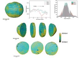
将全场透射 X 射线显微镜(TXM)与 X 射线吸收近边缘结构(XANES)光谱相结合,可获取数十纳米级样品内的二维(2D)或三维(3D)形态和化学特异性信息。这项技术在材料科学和电池研究领域有着广泛的应用。尽管二维 XANES 成像技术具有广泛的适用性,但当样品厚度不均匀时,它也存在信息重叠的缺点。三维 XANES 成像是一种将三维 TXM 与 XANES 相结合的技术,从而能够获取与化学状态有关的三维分布信息。上海同步辐射设施(SSRF)开发了一种三维 XANES 成像方法,并已用于表征商用 LiNixCoyMnzO2(NCM,x + y + z = 1)电池粉末材料的结构和化学状态。成像结果以深度分辨率直观地呈现了颗粒的三维化学状态信息,从而可以直接观察三维镍氧化过程。本文将详细介绍三维 XANES 成像的数据采集、数据处理、量化和可视化分析。
Full-field transmission X-ray microscopy (TXM) in conjunction with X-ray absorption near edge structure (XANES) spectroscopy provides two-dimensional (2D) or three-dimensional (3D) morphological and chemical-specific information within samples at the tens of nanometer scale. This technique has a broad range of applications in materials sciences and battery research. Despite its extensive applicability, 2D XANES imaging is subject to the disadvantage of information overlap when the sample thickness is uneven. 3D XANES imaging combines 3D TXM with XANES to obtain 3D distribution information on chemical states. A 3D XANES imaging method has been established at the Shanghai Synchrotron Radiation Facility (SSRF) and has been used to characterize the structure and chemical state of commercial LiNixCoyMnzO2 (NCM, x + y + z = 1) battery powder materials. The imaging results provide a visual representation of the 3D chemical state information of the particles with depth resolution, allowing for the direct observation of 3D nickel oxidation. This paper will describe in detail the data acquisition, data processing, quantification and visualization analysis of 3D XANES imaging.

 求助内容:
求助内容: 应助结果提醒方式:


